
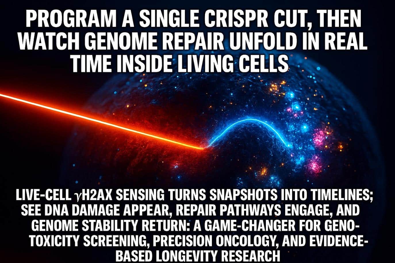
Biologists have long relied on fixed-cell stains that freeze a moment in time, preventing real-time observation. These traditional methods could confirm DNA damage occurred but revealed nothing about how it unfolded. A new live-cell sensor is changing this reality, as described in a Nature Communications study on engineered chromatin readers. The team built a fluorescent reader that recognizes the damage flag γH2AX and lets scientists watch repair occur in living cells with minute-by-minute precision.
The team at Utrecht University reports that the probe binds briefly, releases, and rebinds as damage appears and resolves, a behavior consistent with neutral readouts. According to the Utrecht University news release, those short visits ensure the sensor reports cellular activity without perturbing the native processes, a crucial step for translating bench discoveries into realistic models of cancer therapy, radiation response, and healthy aging.
The Future of Human-Controlled DNA Repair
The analogy for DNA repair is a city’s emergency services; traditional methods provided only a photograph of parked ambulances after a call. The new sensor provides information closer to live traffic data. Researchers can follow where breaks form, how quickly repair crews arrive, and when the streets clear, an advance reflected in Phys.org coverage of the live-cell sensor.
The efficiency of repair is crucial, given that the underlying biology of cellular resilience is strictly time dependent. Repair efficiency interacts with NAD+ metabolism and PARP activity, and the downstream consequences touch familiar ground for Intelligent Living readers, including how telomeres shorten with stress and age and how science is approaching aging as a tractable process, which helps keep longevity discussions grounded.
Key Facts: DNA Repair Sensor Capabilities and Context
- What’s New: A protein sensor built from the MCPH1 tandem BRCT domain binds the damage mark γH2AX in living cells, creating a real-time readout of DNA repair and repair.
- Why it’s Different: The probe shows short residency on chromatin, which reduces the risk of perturbing the very processes it measures.
- Where it Works: Demonstrated in mammalian cell lines and in the animal model C. elegans, enabling both cell culture and in vivo-style observations.
- Why it Matters: Real-Time imaging kinetics can refine genotoxicity screens, inform cancer therapy studies, and align with the broader shift toward single-cell research.
- Contextual Link: The sensor aligns with prior research exploring enzymatic repair of age-related DNA lesions in neurons and what that means for cognition, including an enzymatic pathway that fixes DNA damage.

Engineered Chromatin Readers for γH2AX: The Core Technology
The Signal: What the Sensor Actually Reads
When double-strand breaks occur, cells rapidly tag nearby chromatin by adding a phosphate to the histone H2AX protein, creating γH2AX. The new sensor recognizes this tag and lights up at damage sites, which appear as puncta that grow and fade as repair proceeds.
The Reader: How the Probe was Built
Instead of tagging a repair factor, the team engineered an engineered chromatin reader built from the MCPH1 tandem BRCT domain, a module known to bind γH2AX. By fusing this domain to a fluorescent protein and optimizing linkers, the authors created a minimally intrusive reporter that can be stably expressed in cells for real-time imaging and genome-wide mapping workflows.
The Advantage: Report Without Steering the Outcome
Live reporters often present a challenge: they can subtly alter the very processes they are designed to track. Here, the sensor shows transient binding and rapid exchange on chromatin, so the readout follows the cell’s decisions rather than dictating them. This means researchers can now compare how fast damage resolves under different drugs or radiation doses without the risk of the probe biasing the pathway choice.
The Payoff: From Snapshots to Timelines
Most labs still depend on antibody staining in fixed samples, which delivers a yes-or-no answer after the fact. With real-time imaging, the same cell can be tracked through appearance, peak intensity, and resolution phases. The resulting timeline reveals whether a compound causes damage that lingers, clears quickly, or rebounds. This shift is especially relevant for therapies that rely on DNA repair dynamics, including PARP inhibitors and certain radiation regimens.
Validation Across Models: From Cell Culture to C. Elegans
In Mammalian Cells: Controlled Breaks and Comparable Conditions
The authors show the sensor responding to DNA damage induced by common lab agents and to single, precisely targeted breaks created with CRISPR nucleases. That pairing allows researchers to correlate the location and timing of a defined break with the kinetics of repair seen by the probe, a bridge between genome editing and live-cell DNA repair response.
For readers who follow editing tools, this extends earlier efforts to schedule programmable CRISPR cuts over time into a real-time repair dynamics framework.
In an Animal Model: Watching Repair During Meiosis
The team validated the probe in C. elegans, a transparent roundworm used widely in genetics, marking an important step for organismal studies. That makes it possible to observe repair foci in a living organism and in the specialized context of meiosis, where programmed breaks are part of normal development. Seeing those foci form and resolve in real tissue adds confidence that the sensor can support in vivo-style questions without losing the simplicity of a microscopic readout.
Why this Scale-Up Matters for Health Research
The ability to cross the gap from dish to organism is crucial for studies on cancer therapy response, environmental exposures, and aging biology trajectories. Live kinetics can help differentiate compounds that trigger fleeting stress from those that leave persistent scars, and they provide a cleaner window into how chromatin context influences outcomes across tissues.
Applications: Advancing Cancer Therapy and Aging Biology
Drug and Chemical Safety Testing
Real-time foci appearance and resolution give toxicologists an immediate sense of whether a compound causes DNA damage that is brief, persistent, or cyclical. The high-resolution timeline supports smarter dose finding and flags problematic kinetics early during the discovery phase. The approach also complements population health stories about everyday exposures, including evidence that formaldehyde accelerates stem cell aging, which illustrates the importance of precise readouts of genotoxic stress.
Genome Editing Quality Control
By pairing targeted, single double-strand breaks with real-time imaging, teams can connect where and when a cut occurs with how quickly a cell resolves it. This context significantly improves editing outcomes, especially when researchers need to separate location effects from pathway choice. The framework aligns with controlled perturbation studies such as the CRISPR investigation of DNA repair in space while bringing those ideas into a high-resolution lab setting, and advances in next-generation CRISPR systems with higher precision provide additional control variables.
Radiation and Cancer Therapy Studies
Many anticancer strategies depend on inducing DNA damage that tumor cells cannot tolerate. The new sensor allows investigators to visualize damage formation and clearance kinetics under different radiation regimens or repair-pathway inhibitors. As a biological counterpoint, nature offers protective strategies such as the tardigrade Dsup protein that shields chromatin, highlighting how context can decide survival.
Chromatin Context and Aging Biology
Histone H2AX repair kinetics differ between compact heterochromatin and open euchromatin, helping explain why the same lesion can have different outcomes across cell types and life stages. The DNA damage response relies on cellular energy balance; consequently, longevity discussions often converge on NAD+ metabolism. Nutrient support such as NMN’s role in supporting cellular NAD+ frames why timing and energy status can matter for repair efficiency.
Data Layer for High-Content Screens
The probe’s short residency means researchers can capture many time points without saturating the system. That enables plate-based, high-content screening where the output is not just a single endpoint but a curve that separates fast-resolving hits from slow injuries. These richer kinetics should improve structure–activity relationships and reduce false positives that only appear problematic in static snapshots.

Comparative Analysis: Live Dynamics Versus Outcome Reporters
Live Dynamics Versus Outcome Reporting
Outcome reporters quantify what pathway “won” at a break. The classic Traffic Light Reporter assay reads out homology-directed repair versus end-joining, which is powerful but does not visualize how the decision unfolded over time. In contrast, the live γH2AX sensor shows when damage appears and when it resolves, giving laboratories a view of the journey rather than only the destination.
Probe Classes in Context
Some teams use a γH2AX nanobody approach to follow damage in living cells. While effective in certain models, nanobodies carry different binding and kinetic properties compared to the engineered chromatin reader described here. Others tag repair factors such as 53BP1 to visualize foci, a strategy that can favor or interfere with specific repair-pathway states. As an additional reference point on timing, a live-cell γH2AX kinetics study parsed the distinct early roles of ATM and DNA-PK, underscoring why temporal resolution matters for mechanism.
When Each Method Shines
Choosing the optimal method depends on the specific question: the new live γH2AX sensor is best used when temporal resolution is the primary focus. Here are guidelines for selecting the appropriate approach for DNA repair studies:
- For Spatiotemporal Dynamics: Use the live-cell sensor when you need dynamics in cells or transparent organisms and want minimal interference with pathway choice.
- For Quantitative Tally: Use outcome reporters (like the BLRR) when you need quantitative tallies of HDR and NHEJ across conditions or animal cohorts.
- For Specific Protein State: Use factor-tag strategies when the state of a specific protein is the experimental question and potential perturbation is acceptable.
The new sensor fits neatly into this spectrum by providing the high-resolution timeline that was previously missing, complementing the quantitative outcome reporters.
Future Development: Validation, Scale-Up, and Next Steps
Expression Tuning and Perturbation Checks
Although the study reports short residency and no detectable interference in tested systems, expression level and cell type will always matter. The safest practice is to validate that the reporter does not shift repair timing or pathway choice under your exact conditions before scaling experiments.
Tissue Penetration and In Vivo Readouts
C. elegans validation is an important milestone for organismal biology, and the next challenge is applying in vivo analysis to deeper mammalian tissue. Researchers will likely combine brighter tags, adaptive optics, or tissue clearing with the sensor to extend high-resolution imaging capabilities beyond cell culture.
Standards for Screens and Analytics
High-Content Screening will benefit from common reference protocols and open data formats that encode the full time course. Community benchmarks can include agent panels with known kinetics and parallel readouts from outcome reporters to align dynamics with repair tallies. A foundational review on γH2AX in the DNA damage response provides essential background on why the mark anchors many assay designs, having been a durable biomarker across contexts.

The Live Timeline: Shifting Paradigms for DNA Repair Studies
The shift from fixed slides to live timelines changes what researchers can ask about genome stability. The engineered chromatin reader for γH2AX reports the rise and fall of DNA damage in the same cells and in a simple animal model. This transforms repair from a simple yes-or-no question into a measurable sequence of events. That added dimension should help laboratories design safer compounds, clarify how radiation and repair-pathway drugs interact, and keep aging biology grounded in mechanisms rather than headlines. Most of all, it offers a practical way to see cellular resilience as it happens, which is the first step toward improving it.
Frequently Asked Questions About the Gamma H2AX Sensor
The Mechanism: What The Live-Cell Sensor Binds and Why
It recognizes a phosphate mark on histone H2AX that spreads around a double-strand break. This γH2AX signal acts like a beacon for repair proteins and has long served as a robust biomarker for DNA damage studies.
Potential Perturbation: Does The Probe Interfere with Cellular Repair?
The study emphasizes short, reversible binding and reports no detectable interference in the tested systems, which is consistent with the design goal of reporting without steering. As with any reporter, labs should confirm neutral behavior under their specific expression levels and cell types.
Scale-Up Capacity: Can the Sensor Function in Living Organisms?
The team demonstrates clear foci in C. elegans, a transparent model that is widely used in genetics. This result paves the way for more in-depth tissue research in mammals, utilizing optical strategies that maintain real-time imaging.
Comparative Advantages Over Outcome Reporters
Outcome systems quantify pathway choices, while the live γH2AX reader shows the timing of damage appearance and resolution. Many studies will benefit from pairing both views so that dynamics and final outcomes inform one another.
The Importance of Temporal Resolution in Repair Kinetics
Live imaging experiments show that ATM and DNA-PK contribute differently to early DNA damage signaling, which explains why inhibitor studies can produce distinct temporal signatures.


