
A research team in Denmark and the United Kingdom has built a brain implant that does something no other device of its kind manages: it records brain activity, stimulates neurons with light, and delivers drugs, all through a single fibre thinner than a pencil lead. And it does them at several depths in the brain at the same time.
The device, called the microfluidic Axialtrode, or mAxialtrode, was built by researchers at the Technical University of Denmark, the University of Copenhagen, University College London, and the University of Manchester and published in Advanced Science in a paper titled “Multimodal Layer-Crossing Interrogation of Brain Circuits Enabled by Microfluidic Axialtrodes”. The Technical University of Denmark described the device in February 2026, describing it as one that can illuminate, listen, and deliver medication to the brain.
There is an important caveat to state at the outset, and it is the kind most write-ups skip. This is a research instrument. It has been tested in mice and in laboratory models, not in people. No human trial has begun, and the researchers are still working out whether one is even feasible. Postdoctoral researcher Kunyang Sui, who developed the concept with Associate Professor Christos Markos, says plainly that extensive testing, further development, and regulatory approvals would be needed before the technology could be used clinically.
So this is not a treatment. It is a better instrument for understanding the brain. That distinction matters, and the rest of this article keeps it firmly in place.
What the mAxialtrode Actually Is
The mAxialtrode is a single flexible fibre measuring between 420 and 440 micrometres across. For scale, a human hair is roughly 70 micrometres thick, so the device is about six hairs wide.
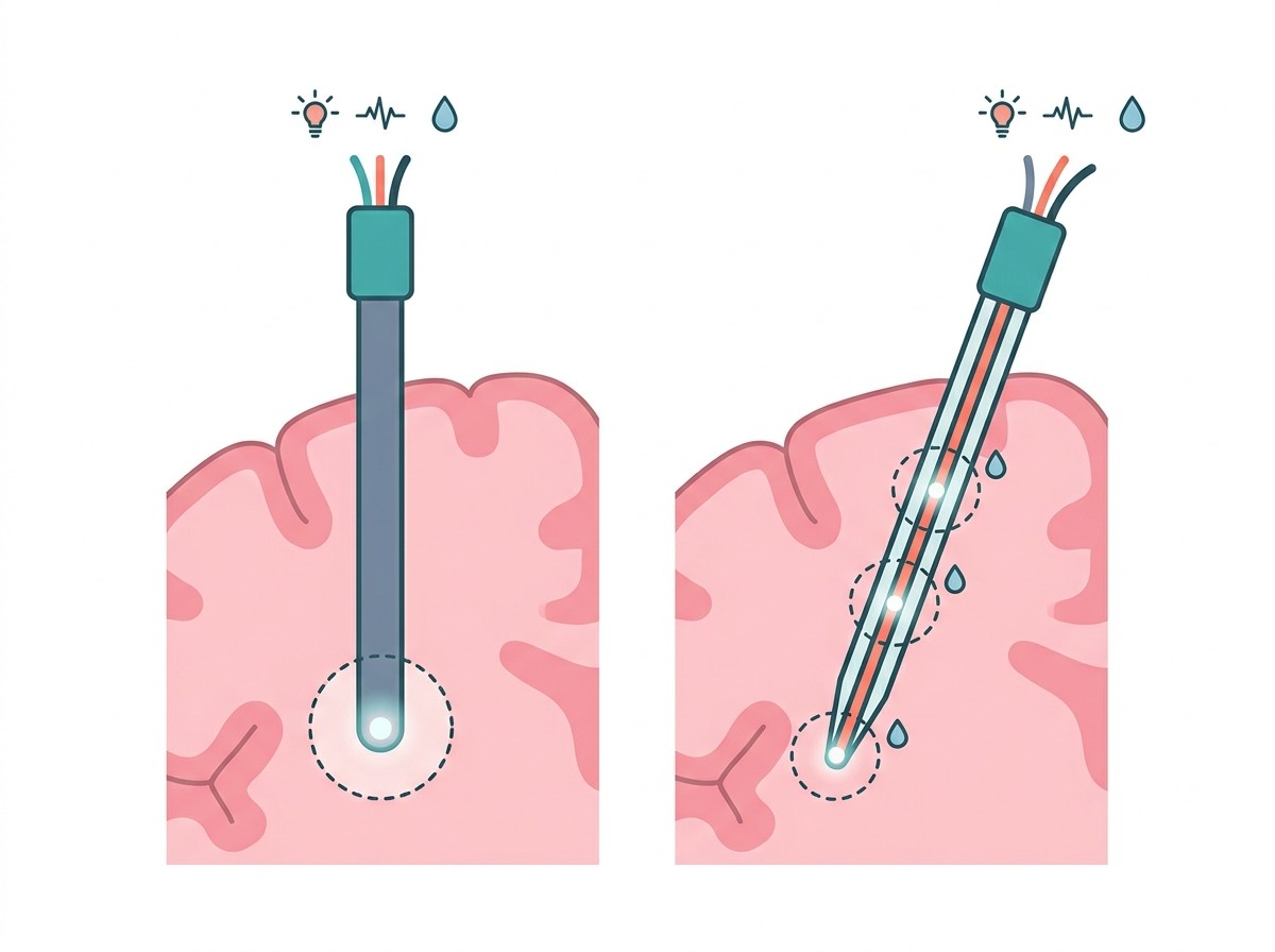
Its architecture is the whole point. A light-guiding core 200 micrometres wide runs down the centre. Around that core sit eight microscopic channels, each about 20 micrometres across, arranged evenly in a ring. Those channels do double duty: they can carry fluid, and they can hold tungsten wires just 20 micrometres thick for measuring electrical activity.
In the mouse experiments, the team used the fibre to do three things at once. They stimulated neurons with both blue light at 470 nanometres and red light at 650 nanometres, the two wavelengths used to drive the most common optogenetic proteins. They recorded electrical activity simultaneously from shallow and deeper brain regions, including the cerebral cortex and the hippocampus, spanning roughly 900 micrometres across the electrode contacts. And they injected different substances at separate depths, with delivery points spaced up to 2.7 millimetres apart.
All of that ran off one lightweight fibre that the mice carried without any obvious signs of discomfort. The fully assembled device weighs between 0.25 and 0.6 grams, depending on which of the two 3D-printed scaffold designs is used.
The device also integrates with a 3D-printed scaffold made from an FDA-approved biocompatible resin, which gives it mechanical stability and keeps it compatible with standard laboratory hardware.
The Problem It Was Built to Solve
To understand why this device is interesting, you need to understand what came before it.
Neuroscientists have long used thin optical fibres to carry light deep into the brain. These fibres power optogenetics, a technique in which specific nerve cells are activated using light. It is one of the most powerful tools in modern neuroscience because it allows researchers to turn particular populations of neurons on and off and watch what happens.
Conventional optical fibres have a structural limitation. They are flat-ended, which means they interact with brain tissue only at the distal tip, the very end of the fibre. All light emission and all tissue contact happen at that single point.
The consequence is that a conventional fibre can stimulate or record from one brain layer at a time. To investigate how signals move between the cortex and the hippocampus, or between a seizure focus and surrounding tissue, a researcher has to insert, measure, remove and reposition. Each insertion wounds tissue. Each repositioning introduces uncertainty about whether the cell population you were watching is the same one you are now watching.
This matters because most brain functions depend on communication between layers. A seizure, for example, is not a local event. It is the spread of abnormal activity through connected circuits. If you can only see one point in that circuit, you are inferring the rest.
That gap is not merely inconvenient. Circuits that span large vertical ranges, such as the seizure networks that connect cortical and hippocampal regions, are exactly the ones a single-point probe handles worst.
Here is the comparison at a glance:
| Feature | Conventional flat-end optical fibre | Silicon-based implants (e.g. deep brain stimulation) | mAxialtrode |
|---|---|---|---|
| Diameter | Varies, typically 100–500 μm | Varies, often 1 mm or more | 420–440 μm |
| Material | Glass or plastic | Silicon, platinum-iridium | Soft polymer composite |
| Active brain layers | One (distal tip only) | Typically one contact site | Multiple, along the length |
| Optical stimulation | Yes | No | Yes (blue and red light) |
| Electrical recording | Usually requires a separate probe | Yes | Yes, via embedded wires |
| Drug delivery | No | No | Yes, 8 channels |
| All three functions in one device | No | No | Yes |
| Approved as a human therapy | No | Yes, decades of use | No |
How It Is Made: The Part Nobody Explains
Here is the detail that most news write-ups about this device skipped, and it is the actual innovation.
The mAxialtrode is not machined, printed or assembled. It is drawn.
Researchers begin with a large polymer rod. They heat it and pull it, in much the same way a thick strand of molten sugar becomes fine thread, except with far greater precision. This is a standard technique in optical fibre manufacturing, and the basic idea is that a thick, bulky preform can be drawn down into an extraordinarily thin, consistent filament.
Getting eight separate fluidic and electrical channels to survive that process, each holding its own wire, without any of them collapsing or merging, is the manufacturing achievement. The channels are not drilled or etched in afterwards. They exist all the way through, from the moment the fibre is formed.
Then comes the trick that gives the device its name: controlled angled cleaving.
After drawing, the fibre is cleaved at a deliberate angle rather than cut flat. Because the cut is angled, the channels are opened and exposed at several different points along the length of the shaft, staggered from one another. Light, fluid and electrical contact can therefore be delivered to multiple depths from a single fibre, instead of only from the tip.
Angled cleaving itself is not new. An earlier generation of thermally drawn multifunctional fibres proposed it as a possible route to depth-specific neuromodulation, but that work never validated the idea functionally, and reaching different depths still required physically advancing the implant between injections. Other approaches relied on mechanically repositioning an inner module. The mAxialtrode is the first to make the angled facet work as an actual functional feature, opening channels and electrodes at different depths so they operate at the same time rather than in sequence.
The name axialtrode comes from this axial redistribution. Function is spread along the length of the device rather than concentrated at a single point. The tip geometry also matters for insertion: an angled tip is mechanically gentler as it passes through tissue than a flat-ended one because it presents a smaller leading profile.
This is why the device is described as needle-thin rather than simply small. The design is not only about diameter. It is about how the fibre enters tissue and how it behaves once it is there.
What Was Actually Tested
It is worth being precise about the evidence, because the published record and the press coverage do not fully match.
Two things happened in the experiments, and they are not equivalent.
In the living mice, the team demonstrated the optical and electrical capabilities. They stimulated nerve cells with blue and red light. They recorded electrical activity from both the cerebral cortex and the hippocampus through the single fibre. The mice carried the implant without any obvious signs of discomfort.
In a brain phantom, a laboratory material engineered to mimic the optical and mechanical properties of brain tissue, they demonstrated the drug delivery. Two coloured compounds were pushed through the uppermost and the lowest channels, producing a delivery span of up to 2.7 millimetres, against 1.27 millimetres for a flat-end fibre given the same volume.
That second point deserves emphasis. The multi-depth drug delivery result is not from a living brain. It comes from a phantom. The device can move fluid to several positions along its length, and the paper’s abstract places drug delivery alongside its in vivo demonstrations, but the experiment behind the 2.7 millimetre figure was benchtop. The paper does not show drugs delivered to multiple depths in a living animal, and it reports no therapeutic outcome.
What the paper does not claim is that the device treats anything. Its own discussion is candid that this is a proof of concept, and the future work it lists, wireless control and real-time sensing among them, is framed as a possibility rather than a plan.
The paper also reports a recording limitation that most press coverage has not mentioned. Light travelling down the fibre produces electrical artefacts on the electrodes nearest the tip, which contaminate the low-frequency part of the recording. The team found this could be filtered out with a high-pass filter at 500 hertz, leaving spike data clean, but it is a real constraint on using the device for anything that depends on slow brain rhythms.
The researchers are now patenting the underlying technology and, according to DTU, clarifying what would be required to test the electrode in a clinical department. That is a very early stage. Nothing has been announced beyond exploration.
Why Soft Implants Matter More Than They Sound
Here is the part of this story that has consequences well beyond one device, and it is the reason the material choice is not a detail.
Brain tissue is extraordinarily soft. Its Young’s modulus, a measure of stiffness, is typically in the range of 1 to 10 kilopascals. Silicon sits at around 100 gigapascals. Platinum is in the region of 100 megapascals.
That is a mismatch of four to five orders of magnitude between a conventional implant and the tissue it sits in.
Why does that matter so much? Because brain tissue moves. Every breath, heartbeat and movement of the body sends tiny mechanical forces through the skull and into the tissue. A stiff implant does not move with the tissue. It repeatedly scrapes and presses against the neurons around it, and micromotion at this scale is a major driver of the injury response.
The consequence is the foreign body response, a multi-stage process that begins when the device is inserted. Blood vessels rupture. The blood-brain barrier is disrupted. Proteins adsorb onto the implant surface. Microglia, the brain’s resident immune cells, activate almost immediately and release inflammatory signals. Astrocytes, the star-shaped cells that help maintain the neural environment, then shift into a reactive state and build a dense layer of fibrous tissue around the implant shaft.
That layer is called a glial scar. It is the same underlying problem researchers have approached from other directions, including efforts to monitor brain activity with microscopic injectable sensors that avoid permanent implantation altogether.
And it is the single biggest reason neural implants degrade. The scar forms an insulating capsule around the device. It pushes the recording sites physically farther from the neurons they are meant to detect. It raises the electrical impedance at the interface. Signal-to-noise ratio falls. Fewer individual neurons can be resolved. The stimulation threshold needed to evoke a response rises.
Over months to years, an implant that worked beautifully on day one can slowly stop working, for reasons that have nothing to do with the electronics and everything to do with biology.
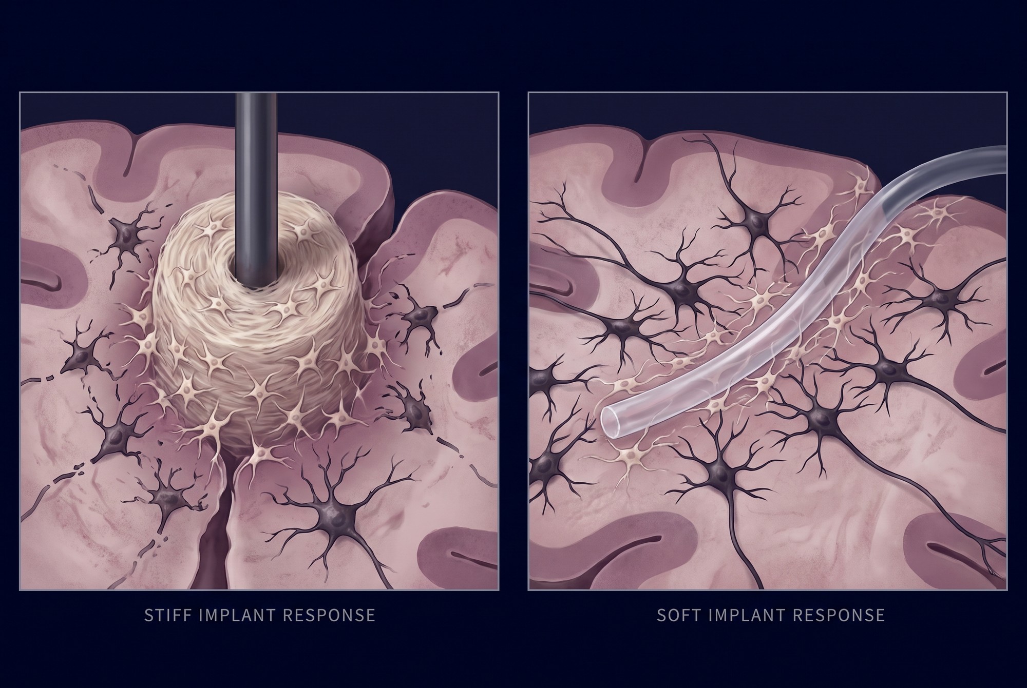
This is the context in which the mAxialtrode’s soft polymer construction stops being a spec-sheet detail. Softness is one of the main strategies the field uses to reduce the mismatch. The paper’s abstract describes the device as significantly suppress[ing] the inflammatory response compared to conventional silica fibers. The experiment behind that statement compared the implant against a flat-end fiber of the same polymer and the same diameter, which suggests most of the measured benefit came from the angled tip and the smaller volume it puts into the tissue rather than from the polymer choice on its own.
Designers are pursuing the same goal from several directions at once. A 2025 review in npj Flexible Electronics catalogues the current strategies for chronically implantable neural probes, covering reduced implant footprint, mechanical softening, porous surfaces that let tissue integrate with the device, and bioactive coatings that release anti-inflammatory drugs to locally suppress the immune response. Each approach attacks the same foreign body response by a different route.
It is also worth being careful about the limits of that finding. The comparison rested on two mice per group, with seven tissue slices analysed per group, four weeks after implantation, and the reported difference fell just short of conventional statistical significance at p = 0.0547. That is an encouraging signal from a small, short study rather than proof of long-term tolerance, and it is not the same as decades of stable function in a human, which is what a chronic therapeutic implant would require. The brain-computer interface field remains candid that chronic biocompatibility, scarring and signal longevity are still unresolved.
What It Could Mean for Epilepsy, and How Far Away That Is
The team points to epilepsy as a motivating application, and the logic is sound. A seizure is a network event, which is exactly the kind of phenomenon that is hard to study with a single-point probe. Being able to record and stimulate across multiple layers simultaneously could help researchers understand how abnormal activity propagates and where it originates.
On the therapeutic side, the researchers describe a scenario in which the device delivers drugs to a precise location while simultaneously applying electrical or light stimulation to a selected area. That is a genuinely interesting combination, because it would allow closed-loop intervention: sense what is happening, then respond at the same site.
But three things stand between this device and a patient’s seizure clinic.
- It has never been in a human. There is no human safety, dose, surgical or long-term data of any kind.
- Optogenetics requires genetic modification. The light-based stimulation only works on neurons that have been engineered to respond to light. In a mouse, researchers can breed or inject animals with this capability. In a person, that opens a completely different category of risk, safety and ethics that has nothing to do with the fiber itself.
- Multi-depth drug delivery has not been shown in a living brain. The 2.7 millimetre result came from a brain phantom. The channels were characterised at safe injection pressures and are intended for virus delivery in optogenetics work, but whether they stay open, deliver a predictable dose and avoid blocking or leaking over months inside a living brain is unknown.
That is a meaningful distance, and it is the honest place to leave the story. The gene-therapy route to neurological disease is being pursued in parallel, and the entry of DREADD-type gene therapies into human trials illustrates how differently the same clinical goal can be approached.
Frequently Asked Questions
Are brain implants possible?
Yes, and some are already in routine clinical use. Deep brain stimulation has been approved and used for decades, most commonly in Parkinson’s disease, essential tremor and dystonia. Responsive neurostimulation is used for focal epilepsy. Cochlear implants restore hearing to hundreds of thousands of people. Spinal cord stimulators manage chronic pain.
What is new here is a specific combination of capabilities in one very small device, not the idea of implanting electronics in the brain.
What are the risks of brain implants?
The main risks are well documented and fall into a few groups.
- Infection and hemorrhage during or after surgery, including bleeding in the brain.
- Foreign body response and glial scarring, which degrade signal quality over time and can leave the device non-functional.
- Infection of the implanted hardware or of the tissue path around it, which can be difficult to treat and sometimes requires hardware removal.
- Device migration or drift, where the probe moves relative to the target neurons and stimulation becomes less precise.
- Hardware failure, including battery depletion for the wireless units used in some devices.
For the mAxialtrode specifically, all of these apply, plus unknowns, because there is no human data whatsoever.
How much would a brain implant cost?
For approved, clinically available devices, the figure is substantial and varies enormously. A cochlear implant, including the device, surgery and rehabilitation, typically runs into tens of thousands of dollars. Deep brain stimulation carries substantial upfront costs for the stimulator, leads and implantation, plus ongoing programming and battery replacement over the life of the device. Both figures vary widely by country and insurer.
There is no cost figure for the mAxialtrode, and any article giving you one is guessing. It is a laboratory instrument in the early stage of being patented.
Is this Elon Musk’s Neuralink?
No. These are unrelated technologies addressing different problems.
Neuralink is pursuing a high-bandwidth brain-computer interface, essentially a very thin array of many electrodes designed to record from large populations of neurons and eventually send signals outward, with an emphasis on restoring function to people with spinal cord injuries. It is focused on channel count and bandwidth.
The mAxialtrode is a single fiber built for laboratory neuroscience, aimed at the opposite constraint: reaching several brain depths at once and combining light, electrical and chemical functions in one device. It is not a communication interface.
That the mAxialtrode would not obviously function as a Neuralink-style device is a limitation rather than a flaw. It was built to answer a different question.
How does this compare to deep brain stimulation?
They solve different problems at different stages.
Deep brain stimulation is an established therapy. A surgically implanted pulse generator delivers electrical stimulation through leads placed at a specific target, often the subthalamic nucleus or globus pallidus. It has three decades of clinical use and adjustable settings.
What DBS generally cannot do is record and stimulate simultaneously at multiple depths, deliver drugs, or use optical stimulation. The mAxialtrode does all three, but in mice.
The honest summary is that DBS is proven and limited, while the mAxialtrode is capable and experimental. They are at opposite ends of a very long development curve.
Conclusion
The most interesting thing about the mAxialtrode is not that it does three things at once. Multifunctional probes have been attempted before. It is that it does three things at once along a single needle-thin fiber, across several depths of brain tissue, by turning an angled cut in a drawn fiber into a working feature. Earlier multifunctional fibers had proposed the idea. None had shown it functioning.
As a research tool, that combination is genuinely valuable. It means fewer insertions, less tissue damage, and the ability to watch how activity moves between layers rather than inferring it from one point.
As a treatment, it is a patent application and a mouse experiment away from a great many things. Kunyang Sui’s own caution is the right frame: this needs extensive testing, further development and approvals before it reaches a clinical department. Anyone reading a version of this story that skips that sentence is reading an abridged version.
What is genuinely encouraging is the direction of travel. If the field’s central unsolved problem is that brain tissue is orders of magnitude softer than the devices placed inside it, then engineering devices that match the tissue is not a workaround. It is the condition for everything that comes next.


