
Every heartbeat, thought, and muscle contraction relies on the precise flow of ions across cellular membranes, confirming the body’s fundamental nature as an electrical organism. Recent scientific advancements pose a provocative challenge: harnessing these natural electrical signals to guide how stem cells grow and develop.
Researchers reported that precisely tuned electrical pulses can nudge stem cells into changing shape and stiffness within minutes, creating physical cues that may determine what type of cell they eventually become. This research, including a recent RMIT-led stem cell stimulation study, demonstrates how to create implants and materials capable of communicating with living tissue in its own electrical language.
The concept of “zapping cells” may sound futuristic, yet it rests upon decades of research confirming that cells inherently respond to small electrical fields. The body naturally generates microcurrents for essential processes, including wound healing, bone repair, and embryonic development.
RMIT’s work adds a real-time look at how living stem cells physically respond to electrical stimulation, providing data that could shape the next generation of tissue engineering and regenerative medicine, alongside early clinical efforts such as a dry age-related macular degeneration trial that aims to rescue light-sensing cells in the eye.

Essential Facts: Bioelectric Stem Cell Stimulation
- Electric fields can guide movement: Mesenchymal stem cells move directionally in electric fields between 25 and 300 millivolts per millimeter, roughly doubling their migration speed compared to unstimulated cells in controlled lab experiments.
- Cells reshape within minutes: The RMIT research team observed stem cells adjusting their stiffness and cytoskeleton structure almost instantly after receiving controlled electrical cues.
- Electricity replaces chemicals: Instead of relying solely on biochemical growth factors, bioelectric stimulation offers a non-chemical, tunable way to influence cell fate.
- It is already used clinically: Bone growth stimulators and wound-healing therapies already rely on similar low-level electric fields to accelerate tissue repair.
- Potential applications: Smart implants, bioreactors, and adaptive biomaterials that “talk” to cells could emerge from this research.
Cells Already “Speak Electricity”—We Are Learning How to Answer
Every living cell maintains a voltage gradient across its membrane, a tiny but powerful difference in charge that drives essential life functions. In skin, this voltage difference is about 10 to 60 millivolts, and when tissue is injured, the electrical field surrounding it changes. These subtle shifts act like signposts, helping nearby cells migrate toward the damaged area. Scientists call this process electrotaxis.
Electrotaxis research demonstrates that bioelectric cues play a key role in wound healing and tissue regeneration. For example, controlled electrical stimulation has been shown to enhance the alignment and growth of cells in lab-grown tissues. These signals help coordinate how cells organize into functional structures, much like a conductor guiding an orchestra.
Translating these natural electrical languages into engineering tools is the next crucial objective. If scientists can learn how to deliver precise pulses that mimic the body’s own patterns, they could potentially train stem cells to form bone, nerve, or muscle tissues without relying entirely on complex chemical cocktails. This vision is what RMIT’s work begins to make possible.

Real-Time Observation: Stem Cells Reshape in Minutes via Electrical Signals
RMIT scientists combined atomic force microscopy (AFM) with computer modeling to observe how human mesenchymal stem cells physically change when exposed to electrical cues in their 2025 cell reorganization study. AFM is a high-resolution imaging technique that measures the stiffness of living cells down to the nanometer. This instrument is an example of advanced microscopy tools in biomedical research. The team found that when stimulated, stem cells reorganized their internal scaffolding, the cytoskeleton, and altered their stiffness within just a few minutes.
Cell stiffness is tightly linked to its biological fate, making this observed change crucial. Softer cells tend to become neurons or fat cells, while stiffer ones lean toward bone or cartilage. By measuring how quickly these shifts occurred, the researchers created a predictive model that could forecast how cells might respond to different electrical patterns. The findings suggest that tailored electrical stimulation could become a precise steering mechanism for tissue growth.
The research remains in the laboratory stage, yet it represents a significant step forward. It shows that bioelectricity is not merely a side effect of biology but a controllable input variable in the design of living materials. The next challenge is to test whether these mechanical and electrical responses can be replicated at the scale of tissues and, eventually, functional organs.
Mechanotransduction: Connecting Electrical Cues to Cell Fate and Destiny
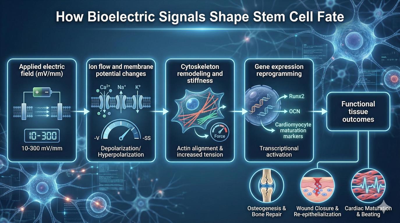
A cell’s shape is more than cosmetic; it helps determine its destiny. When stem cells stretch, compress, or stiffen, they activate internal sensors that send signals to their DNA, switching genes on or off. This process, known as mechanotransduction, converts physical cues into biological instructions. Mechanotransduction helps explain why electrical stimulation has such profound effects: the electrical fields alter the cell’s entire internal tension network, not just the surface.
Researchers have shown that these changes can bias stem cells toward specific lineages. Sustained stiffness, for example, can push cells toward bone formation, while softer mechanical environments favor nerve development. By adjusting the amplitude and duration of electrical signals, scientists may eventually learn to dial in the exact conditions needed for different tissue types.
This mechanical-electrical interplay also explains why studying cell stiffness in real time is crucial. The RMIT team’s AFM approach provides a window into the earliest decision-making moments inside a cell before genes or proteins reveal the outcome. Observing those transitions provides researchers a powerful new tool to understand how life builds itself from the smallest physical motions. This principle is mirrored in bone research, where a newly identified GPR133 bone exercise switch converts mechanical strain into signals that tell skeletal cells to reinforce weakened regions.
In the future, this knowledge could enable engineers to design materials that communicate directly with cells. Imagine smart implants that sense when nearby tissue is under stress and emit microcurrents to stimulate repair, or bioreactors that use electrical cues to precondition stem cells before transplantation. By speaking the same bioelectric language as our bodies, science is learning not to force biology but to collaborate with it.
Integrating Bioelectric Cues in Advanced Tissue Engineering
Using electricity to guide cells is not limited to laboratory tests; the concept could soon shape how tissues are grown for medicine. Electrical stimulation offers a method for aligning, organizing, and maturing cells in three-dimensional environments, complementing 3D bioprinting of living tissues and organs. In engineered tissues, precise electrical patterns might help cells form stronger networks, improving both structure and function.
Electrical pacing protocols have been used, for example, to mature stem cell-created heart tissue, enhancing contraction strength and rhythm. These approaches sit alongside 3D bioprinted organ models and 3D printed human hearts that show how scaffold design and bioelectric cues can be combined. In these systems, electricity acts like a signal booster, guiding communication between cells and the structures they grow on.
The future of tissue engineering may rely on creating environments that are both biologically active and electrically intelligent. By combining bioelectric stimulation with materials science, researchers envision hybrid platforms where microelectrodes deliver tailored pulses to guide cell growth in real time. The objective is to develop tissues that integrate seamlessly into the body and heal faster once implanted.
Proof That Electric Fields Can Guide Stem Cell Behaviors Beyond One Lab
RMIT’s findings are part of a growing global effort to understand how electric fields shape biology. Multiple laboratories have confirmed that human mesenchymal stem cells respond directionally to small voltages in a process known as electrotaxis. Electrotaxis research in 2024 found that cells began to migrate in a coordinated direction at around 25 millivolts per millimeter and nearly doubled their movement speed at 300 millivolts per millimeter.
Other studies demonstrate that electrical stimulation affects gene expression and differentiation. Experiments using conductive nanofiber scaffolds have shown enhanced bone formation markers after several days of exposure to controlled electrical pulses. Research into wound healing further supports these results, because tissues naturally generate electric fields that help close injuries, and artificially applied currents can amplify the effect.
Together, these findings suggest that bioelectricity provides a universal control layer for biological repair and regeneration. Instead of relying solely on chemical growth factors, engineers may soon design devices that use programmed electrical patterns to promote specific cell outcomes.

Immediate Applications: Bone Repair, Wound Healing, and Bioelectronic Devices
Fully engineered organs remain years away, yet electrical stimulation technologies are already finding practical uses. The most immediate applications lie in bone repair, nerve regeneration, and wound healing. Clinical devices, such as FDA bone growth stimulators, use gentle electrical currents to speed up the healing of fractures that struggle to mend on their own. Innovations like ultrasonic 3D printed bone casts also show how mechanical energy can be engineered into bone repair.
Researchers are also exploring electrically conductive dressings that deliver microcurrents to chronic wounds, improving blood flow and encouraging cell migration. This echoes broader advances in next-generation wound-healing medical technologies.
In the laboratory, bioreactors equipped with microelectrodes are being tested to precondition stem cells before transplantation. These devices simulate the body’s electrical environment, training cells to respond more predictably once implanted and complementing regenerative strategies such as stem cell therapy strategies and lab-grown human mini livers. This step could improve the success rate of regenerative therapies while reducing the need for chemical additives.
The next phase will likely combine electrical stimulation with smart materials. Polymers that adjust their conductivity in response to biological signals could create complex feedback loops. This is comparable to bioelectronic blood vessel grafts that integrate biodegradable electronics to sense tissue changes and automatically adapt the electrical environment. Such technology would transform implants from passive objects into active partners in healing.
Major Challenges: Pattern Design, Safety, and Scaling Bioelectric Technology
Despite the promise of bioelectric steering, significant challenges remain. The first significant challenge is pattern design. Researchers must determine which stimulation parameters reliably produce desired outcomes across different cell types. Key elements to define include:
- Electrical Waveforms: Determining the ideal shape of the electrical signal.
- Voltages and Durations: Setting the correct strength and timing for the signal.
Inconsistent results stemming from small variations in signal strength make reproducibility a major obstacle.
Safety and stability are also critical. Long-term exposure to electrical currents risks heat or electrochemical reactions that damage tissues. Consequently, researchers must prove that all stimulation parameters are safe for human use before moving from laboratory to clinic. Scaling these technologies from single-cell experiments to three-dimensional tissues introduces engineering complexities in uniformity and control.
Function matters far more than appearance. It is not enough for cells to grow in the right shape; they must perform the right biological roles, conducting signals, contracting muscles, or producing enzymes. Proving that bioelectric stimulation leads to fully functional tissues will take time, but it is the essential next milestone for the field.
The Future Roadmap: Bioelectric Implants and Advanced Organoid Development
The coming years will reveal whether electrical stimulation can bridge the gap between biology and technology. Researchers are working toward standardized stimulation protocols and closed-loop systems that can adjust signals automatically based on real-time feedback from growing tissues. Advances in machine learning may soon allow scientists to predict the optimal electrical patterns for specific regenerative outcomes.
Another exciting frontier is the development of bioelectronic implants, including tiny implantable wireless devices that are flexible and integrate with the body’s natural circuitry. These could continuously monitor healing and deliver corrective microcurrents when needed. This approach builds on early demonstrations like biodegradable electronic blood vessel graft designs. Studies in organoid development and lab-grown complex human organs are beginning to incorporate electrical patterning to improve the structure and function of lab-grown tissues.
As science refines its understanding of bioelectricity, one thing becomes clear. The future of regenerative medicine will depend as much on physics as it does on biology. Electricity, once viewed as a byproduct of life, is proving to be one of its most powerful tools.
Bioelectric Stem Cells: Common Questions Answered
What exactly happens when stem cells are “zapped”?
The electrical pulses change how ions move across a cell’s membrane, which triggers mechanical shifts within its cytoskeleton. These changes affect stiffness and shape, influencing how the cell differentiates into specialized types.
Does electrical stimulation grow entire organs?
Not yet. The current research demonstrates how to steer cell behavior, not build complex, functional organs. Tissue-scale development requires vascularization, immune compatibility, and precise structural organization that remain active areas of study.
Is it safe for clinical use?
Low-level electrical stimulation is already used in approved medical devices for bone repair and wound healing. However, translating lab-based stimulation protocols to stem cell therapies will require further testing to confirm safety and efficacy.
How does it compare to chemical growth factors?
Electrical cues offer a non-chemical way to direct cells, reducing dependency on costly or unstable biochemical additives. Combining both methods could yield more efficient regenerative outcomes.
What is next for bioelectric research?
Expect rapid progress in combining microelectronics, biomaterials, and artificial intelligence to design adaptive systems that communicate directly with living cells. This cross-disciplinary field will likely define the next decade of regenerative medicine innovation.


