
Planning facial aesthetic changes is fundamentally an operational engineering puzzle. When you alter structural features, you are modifying a three-dimensional framework that has to balance visual proportions with basic biological function.
The biggest mistake is treating facial surgery like a cosmetic quick fix rather than a permanent structural modification. Consulting specialists in rhinoplasty surgery helps you understand how underlying bone and cartilage frameworks support soft tissue over decades. Taking a systematic approach to structural planning ensures your visual results remain functionally sound for life.
Understanding Soft Tissue Envelope Mechanics
Your skin and soft tissue act as an elastic envelope stretched across a rigid bone and cartilage scaffold. If you alter the underlying scaffold without accounting for skin thickness, the visual output rarely matches your expectations.
Thin skin shows every microscopic irregularity in the cartilage underneath, while thick skin conceals fine structural refinements. Surgeons must build internal structural definition specifically adapted to your skin envelope to prevent long-term distortion.
The Costly Mistake of Digital Filter Expectations
A very common and expensive mistake is bringing two-dimensional photo edits to a surgical consultation and expecting an exact match. People make this error because digital editing tools treat facial anatomy like flat pixels rather than dynamic living tissue.
Photographs ignore skin elasticity, cartilage memory, and healing contraction. When patients force a surgeon to over-reduce cartilage to mirror a flat digital edit, they compromise internal structural support, leading to tissue collapse and costly revision surgeries later.
Tracking Real Swelling Timelines and Metrics
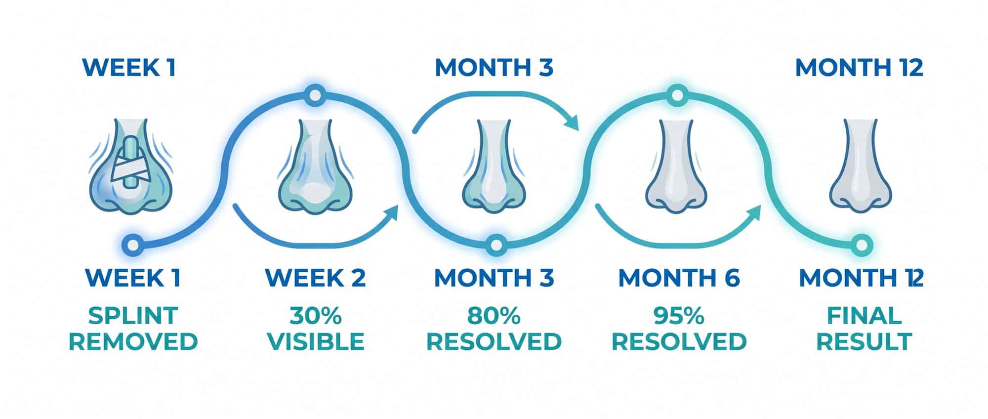
Let us look at a concrete worked example tracking swelling resolution across a full 365-day healing cycle. Many patients panic at week three because early tissue fluid masks structural symmetry.
At week two after splint removal, you see only 30 percent of your final structural contour. By month three, roughly 80 percent of deep fluid drains, but complete skin re-adhesion over refined tip cartilage requires 12 full months. Evaluating your permanent result at month two is like judging a building before the concrete foundation sets.
Skin Envelope Decision Rule for Planning
You do not need a medical degree to understand how your tissue type dictates your surgical plan. You can apply a straightforward decision rule based on skin thickness and elasticity.
If you have thick sebaceous skin, insist on strong structural cartilage grafts to project definition through the dense soft tissue layer. If you have thin delicate skin, prioritise smooth graft camouflage techniques to prevent sharp cartilage edges from showing through over time.
Core Postoperative Operational Controls
Managing your surgical recovery requires strict operational protocols during the critical initial healing phase. Protecting your structural framework while tissue settles prevents minor shifts in alignment.
Focus on these core operational guidelines:
- Maintain head elevation: Sleep at a 45-degree angle for fourteen days to encourage rapid venous fluid drainage.
- Avoid localised pressure: Refrain from wearing heavy glasses directly on structural bones for six full weeks.
- Protect against impact: Pause high-intensity contact sports for three months to prevent displacement of healing bones.
Following these simple rules protects your investment and ensures your underlying framework heals in precise alignment.
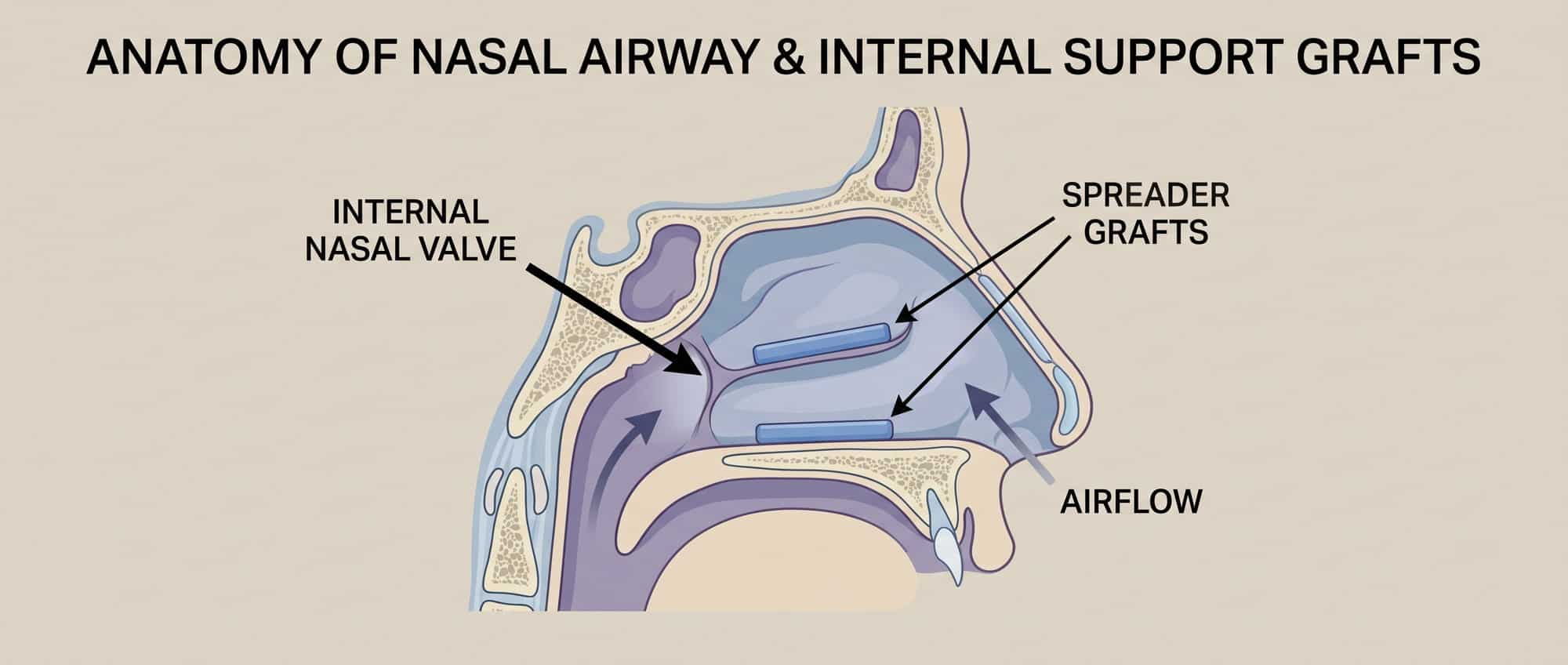
Balancing Airway Function with Visual Symmetry
A successful structural adjustment should never force you to compromise on basic respiratory function. True aesthetic refinement works in harmony with internal airflow mechanics.
When surgeons use internal support grafts like spreader grafts, they reinforce the internal nasal valve while straightening external contours. Prioritising functional airflow alongside visual balance guarantees a result that feels as good as it looks.
Planning facial structural changes is ultimately about treating your face like a complex three-dimensional system. By avoiding the trap of digital photo filters, respecting realistic swelling timelines, and choosing technical approaches tailored to your skin thickness, you ensure your results stand the test of time. Take a structured approach, give your body time to heal, and move forward with quiet confidence in your decision.


