
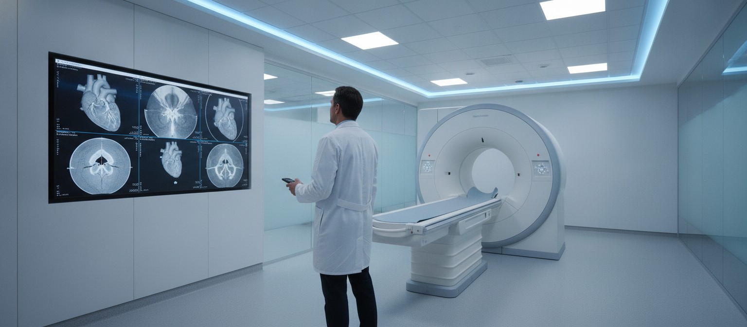
A clinical cardiac magnetic resonance scan generates hundreds of cross-sectional images capturing every contraction of the cardiovascular system. For a radiologist or cardiologist, manually contouring heart chambers to calculate cardiac function typically takes around 13 minutes per patient. A new model for AI heart MRI analysis can perform that exact volumetric assessment in just two to three seconds. More importantly, when evaluated across thousands of patients, the algorithm predicted long-term survival slightly better than the measurements logged by attending healthcare professionals.
Measuring the Pumping Heart: AI Versus Clinical Workflow
When doctors evaluate heart failure or assess cardiac risk, their primary metric is often left ventricular ejection fraction. Ejection fraction represents the percentage of blood pumped out of the heart’s main chamber with each contraction. A normal measurement hovers between 50% and 70%, while lower percentages indicate a damaged or weakening heart muscle. Obtaining this number requires drawing outlines around the endocardium across multiple cross-sectional slices at both peak filling and peak contraction.
Because manual contouring is tedious and subject to subtle variation between observers, clinicians often rely on rapid visual estimates during busy shifts. The automated deep learning pipeline evaluates entire multi-slice sequences with strict mathematical consistency.
A 27,000-Patient Study Compares Long-Term Predictions
To evaluate whether automated measurements translate into meaningful prognostic utility, researchers conducted a large UCL-led multicenter study analyzing real-world hospital data. The team examined 27,382 cardiac MRI examinations conducted between 2014 and 2019 across UK and US centers, drawing primarily from the Barts Heart Centre clinical cohort.
The investigators followed these patients over a median span of more than five years, during which 3,458 deaths were recorded. As reported in The Lancet Digital Health in November 2026, researchers compared the predictive accuracy of the automated measurements against the original clinical reports filed by doctors.
Accuracy was quantified using the concordance index, or C-index, where 0.5 denotes random chance and 1.0 reflects flawless survival ranking. The automated ejection fraction achieved 0.669 versus 0.637 for standard clinical reports. Model-fit statistics favored AI strongly (Akaike difference of 161 points), and adding AI to clinician readings improved prediction far more than the reverse. After adjustment for age and sex, the gap narrowed to a C-index difference of 0.004, a small but significant edge. The tighter automated boundaries removed minor manual inconsistencies that can obscure subtle risk differences.
Why Human Verification Remains Essential
Despite the algorithm’s statistical edge and rapid processing speed, the study highlights critical nuances that prevent full diagnostic automation. The research team noted several practical boundaries and caveats:
- Routine clinician oversight: The automated system produced an error rate of approximately 1%, usually triggered by imaging artifacts, arrhythmia-induced slice misalignment, or atypical cardiac anatomy that requires expert manual correction.
- Retrospective study design: The dataset observed historic records collected between 2014 and 2019 rather than testing real-time clinical intervention in an active, prospective clinical trial.
- Marginal statistical gains: While the difference in C-index (0.669 versus 0.637) was statistically significant, the net prognostic improvement became marginal once models adjusted for routine patient risk factors such as age, sex, and underlying comorbidities.
- Unproven patient outcomes: Demonstrating that an automated calculation correlates more tightly with mortality in archival records does not prove that adopting the algorithm will improve clinical decisions or extend patient lives.
Machine Learning Across Cardiovascular Diagnostics
The emergence of automated scan contouring reflects a broader integration of deep learning across cardiovascular care. Automated algorithms are increasingly deployed upstream to identify early risk before patients ever enter an MRI scanner, such as screening for cardiomyopathy using an AI ECG smartphone sensor in outpatient settings.
When patients do require advanced imaging, offloading routine slice contouring to algorithms can alleviate substantial hospital backlogs. A reduction in processing time from 13 minutes to three seconds per scan could allow imaging departments to report on urgent studies sooner and reduce waiting lists.
However, technology functions best as an assistant rather than an autonomous decision-maker. By filtering out the mechanical burden of tracing cardiac borders, radiologists and cardiologists can dedicate more time to examining complex tissue characteristics, evaluating myocardial scarring, and tailoring treatment plans for vulnerable patients.


