
Operative hysteroscopy requires more than a skilled clinician and the right equipment. It depends on a coordinated workflow that protects patient safety, supports clear visualization, and provides everyone in the room with a shared understanding of the plan. Whether the case involves diagnosis, adhesions, fibroids, or uterine polyp removal, a reliable process can help teams work deliberately when conditions change.
The most effective workflow is not simply the fastest one. It is a repeatable approach that combines preparation, assigned responsibilities, fluid awareness, specimen management, and clear follow-up. Hysteroscopy can be performed in an office, ambulatory setting, or operating room when the patient, procedure, staffing, and available resources are appropriate.
Why Hysteroscopy Workflow Matters
A well-organized process reduces avoidable interruptions without skipping essential safety checks. Room setup, equipment testing, fluid tracking, documentation, and communication are closely connected. If a device is unavailable, a fluid total is unclear, or the next clinical step has not been stated aloud, the team may lose valuable time or proceed without complete situational awareness.
For suitable patients, hysteroscopy may allow direct evaluation and treatment during the same encounter. A recent review of office-based operative hysteroscopy highlights how patient selection, clinician training, pain management, and equipment planning influence whether this approach is practical and appropriate.
Steps To Take Before The Procedure
A brief, consistent pre-procedure discussion helps the team start with the same expectations. Before the patient enters the room or before the procedure begins, confirm the following:
- The patient’s identity, planned procedure, indication, consent, and anticipated treatment.
- Relevant imaging, prior uterine procedures, allergies, medications, anesthesia needs, and health conditions that could affect planning.
- The availability and function of the hysteroscope, light source, camera, distension system, suction, energy equipment, tissue collection supplies, and backup instruments.
- How fluid input, output, pressure, and deficit will be measured and announced.
- Potential findings, escalation options, and conditions that would justify pausing, stopping, or changing the plan.
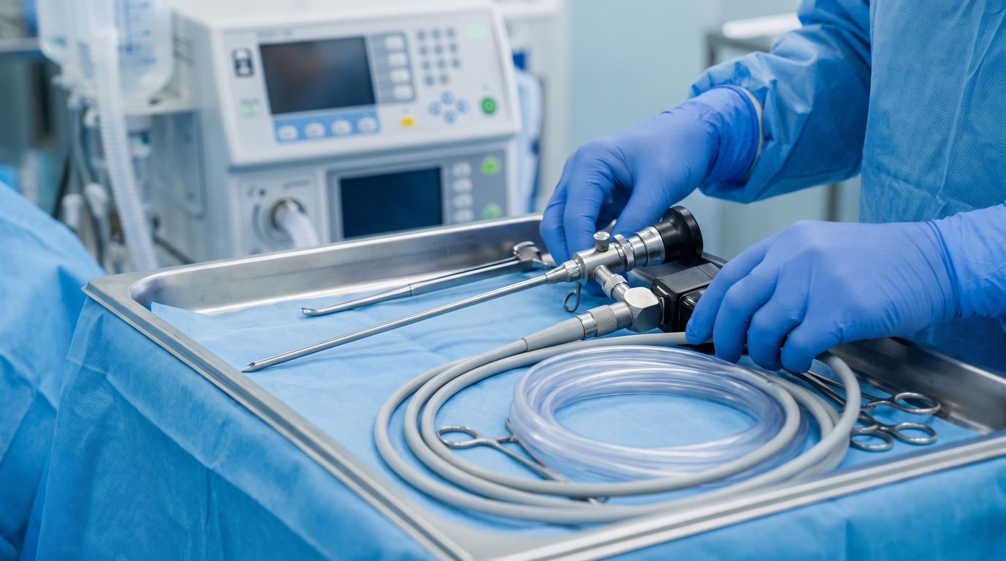
How Each Team Member Supports Safe Care
Clear role assignment prevents important tasks from becoming assumed tasks. The physician leads the clinical assessment and procedural decisions. The circulating nurse can coordinate documentation, supplies, timing, and communication beyond the sterile field. The surgical technologist prepares equipment, maintains sterile technique, and anticipates instrument needs. The anesthesia professional monitors the patient’s comfort and physiologic status while helping the team respond to concerns that affect procedural safety. Coordinators and administrators can support the process by reducing scheduling gaps, maintaining inventory, and ensuring equipment service plans are current.
Managing Visualization and Distension Fluid
Visualization is central to safe hysteroscopy. Distension media creates the working space needed to inspect the uterine cavity and perform treatment, but fluid use must be monitored throughout the case. The team should use a shared process for recording inflow and outflow, calculating the deficit, and repeating critical numbers at planned intervals or whenever conditions change.
Pressure settings, procedure duration, fluid type, bleeding, tissue characteristics, and the patient’s health status can all affect the clinical picture. A designated person should own the fluid-tracking task, while the entire team remains prepared to reassess if the deficit rises, visibility worsens, or the patient’s condition changes. A clear verbal update is more reliable than a number recorded silently on a screen.
Planning Tissue Removal With Greater Precision
Before removing tissue, the operator should assess its location, size, shape, vascular appearance, and suspected depth. Polyps, submucosal fibroids, adhesions, and other intrauterine findings may require different instruments and different sequencing. Planning the order of resection, aspiration, inspection, and hemostasis helps maintain procedural control.
Specimen handling should be discussed before tissue is collected. The team should know which material will be sent to pathology, how containers will be labeled, and how the collection process will be documented. If anatomy becomes uncertain, bleeding increases, fluid values become concerning, or visualization falls, pausing is appropriate. Completing every planned step is not more important than maintaining safe conditions.
Building Better Intraoperative Communication
Short, consistent callouts can reduce ambiguity during active portions of the procedure. Useful communication habits include:
- Stating the current procedural step before changing instruments or technique.
- Announcing meaningful changes in visualization, bleeding, pressure, or fluid balance.
- Repeating critical fluid totals so the information is heard and acknowledged.
- Using a clear stop signal when reassessment is needed.
- Confirming the next step before introducing energy, changing equipment, or ending the case.
Where Technology Fits
Technology can support, but not replace, clinical judgment. Improved imaging, digital documentation, fluid-monitoring systems, and tissue-removal devices may help teams organize information and perform tasks more consistently. Facilities should consider staff training, maintenance needs, compatibility with existing systems, and the clinical value of a new tool before adopting it.
Computer-assisted image review is also an emerging area. Research on the automated classification of endometrial lesions from hysteroscopic images shows that deep learning models are being studied for image interpretation. These tools remain supportive technologies, and trained clinicians must continue to interpret findings in the context of the individual patient.
Post-Procedure Documentation and Follow-Up
The workflow continues after the hysteroscope is removed. Documentation should clearly record the indication, findings, treatment performed, specimens collected, fluid measurements, complications, and any reason the procedure was modified or stopped. Patients need understandable instructions about expected cramping or bleeding, activity, warning signs, pain management, and follow-up timing.
A defined process for reviewing pathology results is equally important. Short team debriefs can identify recurring delays, missing supplies, unclear handoffs, or communication gaps that should be addressed before the next case.

Final Takeaway
Stronger hysteroscopy workflows stem from practical preparation, shared accountability, careful fluid monitoring, purposeful communication, thoughtful use of technology, and reliable follow-up. When every team member knows what to do, what to report, and when to speak up, efficiency can serve its proper purpose: safer, more patient-centered care.


