
A patient walks into a clinical space with a persistent blue tint beneath their eyes. They usually assume it is a lingering bruise. Weeks pass, and the color remains completely unchanged. This is not a hematoma; this is the Tyndall effect. The aesthetic industry has wrestled with this specific optical complication for decades. It manifests as a stubborn blue discoloration after filler placement. Practitioners historically tried to hide the product by injecting it deep into the facial planes. Bury the gel deep enough, and the light cannot reach it. That strategy fails entirely when a patient needs volume directly beneath the epidermal layer. You cannot correct a superficial rhytid with a bolus resting on the periosteum.
A fundamental shift in hyaluronic acid filler technology was necessary to treat these shallow anatomical zones more safely. Medical professionals required a material that could sit near the surface without reflecting the room lights back into the eyes of casual observers. The early generations of dermal implants, like bovine collagen, were not typically associated with this particular blue tint. They presented entirely different challenges, primarily involving allergic reactions and very short longevity. The transition to synthetic hyaluronic acid solved the allergy problem but introduced new optical hurdles. Transparent gels behave like prisms when placed incorrectly. The market needed a structural solution.
The Physics of Short Wavelengths
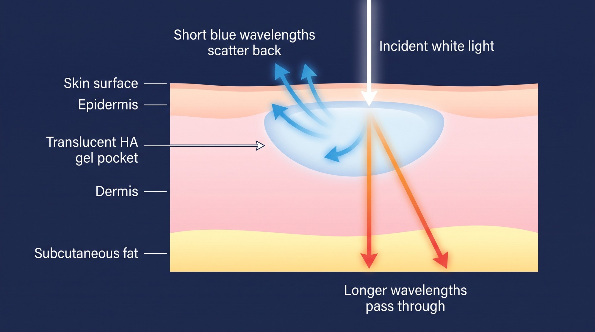
The physics behind this visual distortion are relatively straightforward. The phenomenon revolves around light scattering in the skin. Sunlight and artificial indoor lighting hit the translucent pocket of clear hyaluronic acid resting in the tissue. Red and yellow wavelengths travel straight through the gel because they are longer. Deep tissues absorb these warmer colors. Blue wavelengths are much shorter. They strike the physical boundaries of the gel pocket, scatter intensely, and bounce back toward the surface. The human eye perceives this scattered light as a distinct, unnatural blue hue. Consensus guidance on managing the complication notes that blue light is scattered roughly ten times more than red when it passes through very small particles, and that the effect is more common with particulate fillers. The consensus paper is available in full through PubMed Central. Its authors also stress that injection depth, small aliquot size and product choice are the primary preventive measures.
The local anatomy compounds the problem in thin-skin areas such as the tear trough and the periorbital region. These regions lack sufficient dermal thickness to block or absorb the scattered blue light. Any superficial filler placement in these zones carries a meaningful risk of optical failure if the material does not behave correctly. Place a standard biphasic gel here, and the patient has a considerably higher chance of developing that blue tint. The physical properties of the skin in the tear trough are unforgiving. The dermis is fragile, the subcutaneous fat is minimal, and the underlying vascular network is highly visible. Injectors cannot rely on tissue camouflage in this area. The injected material needs intrinsic properties that limit light refraction.
Product Focus and Material Behavior
Material scientists approached the problem through the structure of the gel itself rather than through injection depth alone. A hyaluronic acid filler built on cohesive polydensified matrix (CPM) technology is formulated to integrate into the dermal layers rather than pushing the surrounding structures apart. Because the gel distributes through the tight interstitial spaces of the skin, it is used in areas where a discrete implant would be visible. Belotero is one of the products formulated this way, and it is usually discussed as the reference example of the category.
The gel behaves as a continuous web rather than as a collection of separate bodies. It conforms to the native collagen and elastin instead of sitting as a firm, isolated water balloon, and clinicians report that this behavior makes delicate areas easier to treat without creating visible lumps or blue shadows. Distribution of CPM gel is handled through licensed medical suppliers, which is also how practices order Belotero fillers for your clinic.
The Chemistry of the Matrix
The manufacturing process behind this behavior is where the technology differs from conventional cross-linking. Many gels are made by cross-linking hyaluronic acid into a bulk block and then reducing it to particles of a chosen size. CPM technology instead builds a hydrogel in which the degree of cross-linking varies throughout the material, so the finished product contains regions of different density rather than a single uniform structure. The cross-linking agent used in hyaluronic acid fillers is most often BDDE, or 1,4-butanediol diglycidyl ether.
The result is high-density zones and low-density zones occurring simultaneously within the same syringe. This variable density underlies the cohesivity and tissue integration that distinguish the material clinically. The high-density sections provide the structural support needed to smooth a stubborn wrinkle. The low-density sections act more like a fluid gel. This softer component allows the material to mold around the patient’s existing tissue architecture. The gel stretches under mechanical stress without snapping or breaking apart. Facial movement requires this type of dynamic flexibility. A rigid gel can eventually look unnatural when the patient smiles or speaks. The polydensified matrix moves with the surrounding muscle and fat.
Rheology and Physical Traits
Filler particle size and gel structure help predict clinical behavior. Biphasic fillers are typically made by passing cross-linked gel through a sizing screen, which leaves distinct particles, and those particles sit under the skin as tiny clear bodies with defined edges. How different dermal fillers work depends heavily on such manufacturing choices. The CPM process instead produces a low-viscosity, soft gel without rigid, distinct particles. The material is monophasic and cohesive, so there are fewer internal boundaries to catch and redirect light, and less of the isolated pooling that makes a clear gel visible through the skin.

Standard rheological testing highlights a few operational traits of this matrix:
- Variable density cross-linking allows the matrix to elongate under shear stress without separating.
- High internal adhesion is intended to keep the product from pooling or migrating away from the injection site.
- Soft outer margins limit the formation of harsh internal boundaries that catch and reflect light.
These physical metrics shape how the product performs when squeezed through a fine needle. Extrusion force tends to be low and smooth. The injector meets less resistance, which supports precision during delivery.
Clinical Application and Delivery
This rheological profile changes the practical rules for superficial dermal and subdermal injection. Dense fillers placed immediately under the epidermis create a visible, firm bleb. Soft cohesive gels make fine-line and skin-quality treatment at shallow depth more feasible. Etched-in wrinkles, such as perioral smoker lines or necklace lines, require a product that can be injected at an extremely shallow depth.
The blanching technique illustrates this clinical requirement. Clinicians insert a very fine gauge needle into the upper dermis until the bevel is barely submerged. They deposit a tiny micro-droplet of the product. The skin turns pale or white for a brief moment as the fluid displaces local blood, then the natural tissue color returns as the gel spreads. A firmer filler can leave a longer-lasting visible bump at the same depth. The cohesive matrix disperses among the local dermal fibers instead.
The choice of instruments interacts with these physical properties. The ongoing debate over cannula versus needle placement depends on the rheology of the specific product. Cannulas work well for deep, broad distribution in the sub-SMAS fat pads. Needles provide the precision required for micro-droplet delivery in the superficial dermis. Injection depth and technique dictate the final visual outcome, and the physical limits of the gel determine which technique is possible.
Reversibility and Safety Protocols
Anatomical variation exists in clinical practice regardless of injector skill, and the ability to reverse a treatment remains a core safety factor in aesthetic medicine. Hyaluronidase hydrolyses hyaluronic acid and is the accepted way to dissolve an unwanted HA deposit, including a superficial one responsible for a blue tint.
How quickly a given gel dissolves has been studied extensively, and the results are not consistent between products or between studies. Some comparisons have found CPM gels comparatively resistant to hyaluronidase, while later work has found the opposite, and dissolution rate appears to depend on hyaluronic acid concentration, cross-linking density and the testing method used. Dense, heavily cross-linked fillers generally need more enzyme or repeat sessions than softer gels. Practitioners therefore treat reversal as a dose-titrated process rather than a fixed protocol. Because a monophasic gel has no separate particle compartments, there are fewer discrete deposits to reach, but the response still has to be assessed in the individual patient.
Tissue mechanics and optical physics remain complex subjects for the medical community. Advanced filler formulation offers partial answers to these persistent clinical challenges. Material design can reduce the optical risk of superficial placement, but it does not remove it. Injection depth, aliquot size and product selection all still determine whether a treated area reads as natural skin or as a visible implant.

