
The physical landscape of the human face changes over time through mechanical failure. Bone resorbs and retreats inward over the decades. Deep fat pads shrink and migrate downward under the skin. The retaining ligaments stretch and lose their baseline tension. The structural support network essentially loses its physical integrity. Correcting this problem requires materials that can closely replicate the mechanical properties of youthful human tissue.
Hyaluronic acid (HA) fillers are widely used to address this biological deficit, and the science of volume restoration rests largely on applied physics. You cannot simply inject a generic liquid into the face and expect it to hold a sharp contour against muscle movement and the constant downward pull of gravity. The implanted material must possess very specific mechanical properties to survive in a dynamic environment. Researchers evaluate hyaluronic acid formulations for cohesivity and viscoelasticity to determine how a gel will behave once placed inside a living host. The goal is to build an invisible, synthetic scaffold that moves like real tissue.
Designing the Structural Matrix
Chemists build HA fillers by binding individual chains of hyaluronic acid together into a three-dimensional matrix. A single syringe is only one point on a much wider spectrum of gel designs, and commercial HA collections include products spanning a range of firmness, cohesivity, and flexibility profiles. Juvederm, for example, is a family of cross-linked HA gels available in several densities, from softer gels used in superficial lines to firmer gels placed deeper against bone. Clinicians and practices that use these formulations source them through a professional Juvederm filler supply.
What matters physically is not the brand but the manufacturing choices behind a given gel. Two syringes of identical HA concentration can behave quite differently depending on cross-linking density, particle size and hydration, and those differences decide whether a gel resists high-pressure forces deep against the bone or moves fluidly with superficial muscle contractions.
The Parameters of Viscoelasticity
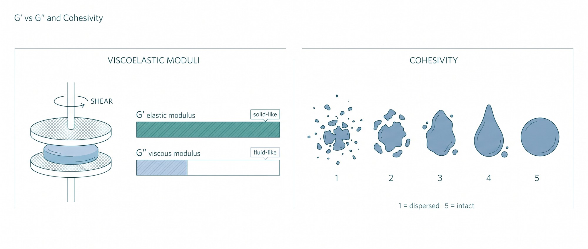
The term “viscoelasticity” defines how a material acts simultaneously as a viscous fluid and an elastic solid. Fluid components allow the substance to flow smoothly out of a needle and conform to a tight space inside the face. Solid components allow the substance to resist deformation and hold its shape over time against external pressure. Rheological testing is the standard laboratory method used to quantify these characteristics in any HA filler formulation. Scientists place the experimental gel between two plates inside a rheometer and apply rotational shear forces to measure the mechanical response in real time. The equipment measures the force required to deform the gel and records how the gel recovers when that force is removed. The procedure allows scientists to map the mechanical profile of the filler mathematically.
This testing produces two primary metrics. The elastic modulus (G-prime) measures the gel’s ability to return to its original shape after a force is applied and released. A high G-prime means the gel is firmer and holds its shape better under shear stress. The viscous modulus (G-double-prime) represents the fluid-like behavior of the formulation. It measures how much of the applied deformation energy the gel dissipates in the process. A high G-double-prime indicates a softer, more fluid material that spreads easily through tight spaces.
Both properties have to work in balance. A filler with an extremely high elastic modulus and almost no viscous component would feel rigid under the skin. A filler dominated by its viscous modulus would behave more like a syrup, flattening out and migrating away from the injection site as soon as a muscle moved.
The Binding Force of Cohesivity
Cohesivity is a separate physical property. It measures the internal adhesive forces binding the gel matrix together, or the material’s reluctance to dissociate and break apart when subjected to dynamic compression. A highly cohesive gel stays intact as a single, unified mass. A gel with very low cohesivity tends to disperse, fracture, and integrate rapidly into the surrounding microscopic tissue spaces.
A 2015 clinical review published in the journal Plastic and Reconstructive Surgery introduced a standardized visual assay for cohesivity and used it, alongside elasticity and viscosity measurements, to characterize six U.S. Food and Drug Administration–approved fillers. The authors positioned cohesivity as a property that complements G-prime rather than mirroring it: gels that are similar in elasticity can still differ in how intact they remain when extruded and dispersed.
That difference matters under load. A very firm gel with low cohesivity may fracture and split apart under the repetitive movement of the orbicularis oris muscle around the mouth. A softer gel with high cohesivity will stretch, flatten, and adapt to the movement while keeping its internal structure. Over time, this mechanical interplay dictates the durability of the implanted volume. When a filler lacks appropriate cohesivity for a highly mobile area, the forces of the facial muscles can degrade the gel matrix prematurely, and the correction fades faster than intended.
Engineering the Gel Chemistry
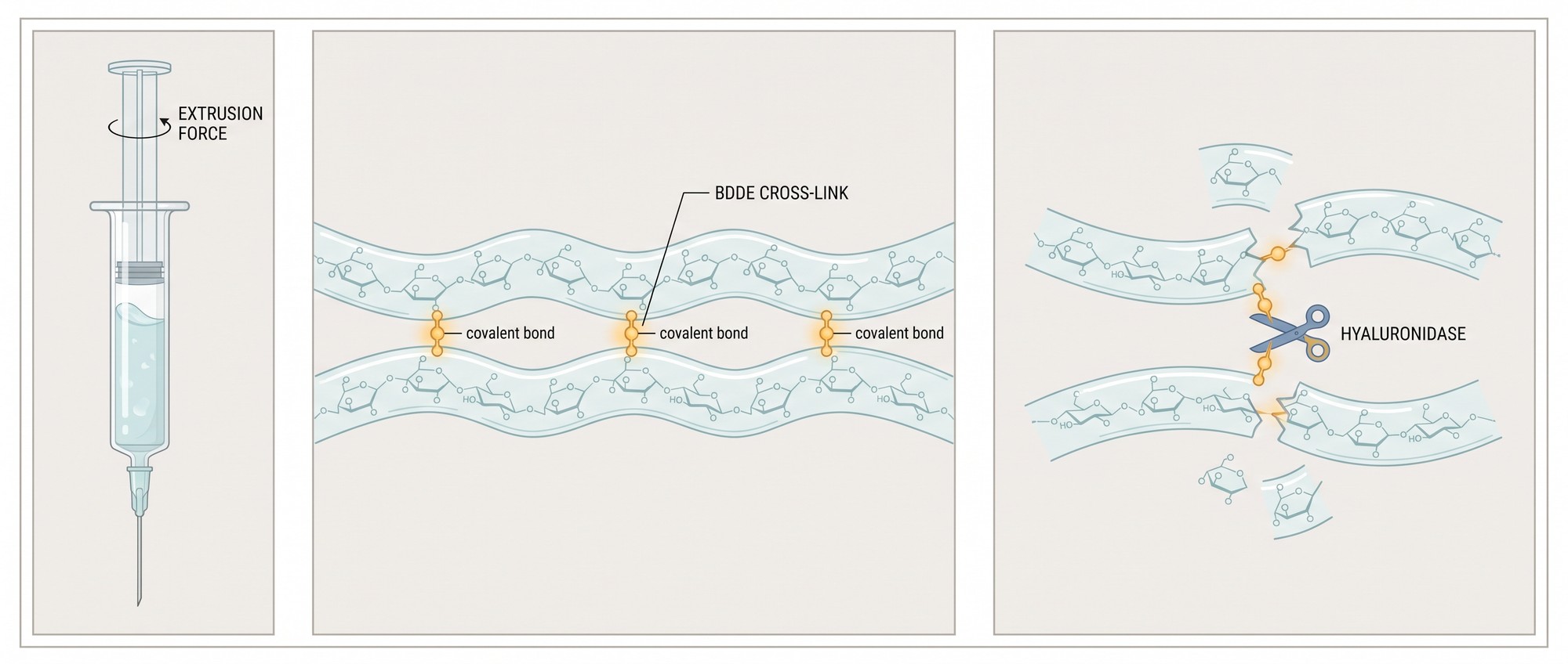
Manufacturers manipulate several variables in the laboratory to achieve the desired physical properties for different anatomical targets. Cross-linking is the foundation of the process. Native, uncrosslinked hyaluronic acid has a short residence time in the body, measured in hours to a few days, because hyaluronidase and reactive oxygen species break it down. Chemists use binding agents such as 1,4-butanediol diglycidyl ether (BDDE), the cross-linker used in most currently available HA fillers, to create covalent bonds between HA chains. This network resists enzymatic degradation and transforms a liquid solution into a semi-solid gel.
The chemistry here is delicate. If the bonds are too loose, the filler degrades rapidly. If the bonds are too tight, the material becomes difficult to work with. The cross-linker acts as a bridge between strands of hyaluronic acid, locking the sugar molecules into a fixed geometric pattern that the body does not recognize immediately. It takes longer for hyaluronidase to break down a densely cross-linked matrix than free-floating hyaluronic acid.
The degree of cross-linking directly influences the behavior of the final product. An increase in cross-links usually raises the elastic modulus, making the gel stiffer and more resistant to mechanical stress. But bonds cannot be added indefinitely. Excessive chemical modification can render a gel brittle or affect its biocompatibility in host tissue.
HA concentration and gel hardness also share a relationship. A higher total milligram count of hyaluronic acid per milliliter of fluid generally produces a stiffer end product. This stiffness contributes to the material’s lift capacity, meaning how efficiently the implanted gel pushes upward against overlying tissue and resists the downward pull of gravity. Deep skeletal volume loss requires a product with high lift capacity to simulate the support normally provided by thick bone and dense deep fat pads.
Hydration Dynamics and Structural Integration
Hyaluronic acid is inherently hydrophilic. The gel swelling factor describes how much ambient fluid the filler absorbs after it enters the biological environment. Gels formulated with little free water in the syringe draw moisture from the surrounding tissue, and that influx of water can produce swelling that appears some time after placement rather than immediately. Published water uptake measurements show the spread is wide: in one comparative laboratory study of 23 fillers, water uptake factors ranged from roughly 2.5 for a non–animal-stabilized gel to more than 9 for a highly cross-linked formulation. In vitro uptake does not translate directly into clinical swelling, but the direction of the relationship holds.
Formulators account for this osmotic pressure to limit unwanted overcorrection. A gel intended for the thin-skinned under-eye area requires a low swelling factor. The thermodynamics drive the behavior: water molecules move toward areas of higher solute concentration, and a densely packed gel behaves like a microscopic sponge. The degree of osmotic pull depends on how the gel was treated before it went into the syringe. Fully hydrated gels have little room to take up more water. Partially hydrated gels expand as they equilibrate, and a review of in vivo volume change describes the injected volume peaking at roughly 1.8 times the original after about four weeks before gradually declining.
The physical architecture of the gel matters as much as its chemistry. Particle size and gel structure determine how the material interacts with host biology. Some manufacturing methods create granular, biphasic gels containing distinct, uniformly sized particles suspended in a liquid carrier. Others produce smooth, monophasic matrices with no distinct particles.
The size of the particles or the density of the matrix strongly affects extrusion force and injectability. A thick, highly cross-linked gel requires substantial pressure to push through a fine 23- to 27-gauge needle, and extrusion forces measured across commercial fillers fall in a range of roughly 12 to 20 newtons. Manufacturers sometimes include a small percentage of uncross-linked HA to act as a mechanical lubricant, lowering the extrusion force and allowing the material to be deposited without excessive hand fatigue. Pushing a non-Newtonian fluid through a narrow cylinder requires the force applied to the plunger to overcome both the internal resistance of the gel and friction against the needle wall. A drop in viscosity under shear stress lets thick gels flow temporarily, and the gel regains its structured state once it leaves the needle tip and the stress is removed.
Navigating the Tissue Planes
Once the gel leaves the tip of the needle, it faces the physical realities of human anatomy. Tissue integration and spread dictate whether the final contour looks natural or artificial. The gel must navigate through fascial layers, muscle fibers, and fat compartments, and the anatomical layer dictates the physical requirement. A filler placed directly over a highly active muscle group experiences constant shearing forces and needs high cohesivity to survive them. A filler placed in a static fat pad experiences little shearing but bears the weight of the tissue above it, so it needs a high elastic modulus.
Formulations handle that journey in different ways:
- A high-G-prime, highly cohesive gel placed deep on the periosteum stays where it is placed, lifting the muscle and fat above it like a small tent pole.
- A low-G-prime, low-cohesivity gel placed superficially in the dermis spreads laterally, intertwining with existing collagen fibers to smooth fine lines without creating lumps.
The mechanical demands of the face vary by the millimeter. The chin requires projection and rigidity. The lips require flexibility and softness. Scientists manipulate the core physics of the gel to create different tools for different anatomical problems. Rheological data tells us how the material will behave long before it touches living tissue, which replaces guesswork with measurable physical properties when rebuilding the structural foundation of the face.

