
Cartilage does not operate in a vacuum. It sits within an isolated anatomical capsule, completely dependent on the surrounding fluid for nutrition and physical protection. The environment inside a healthy joint relies on a highly specific biological balance, a baseline state known as synovial fluid homeostasis. Disruption here leads directly to cartilage breakdown. A cascading series of destructive events degrades the protective barrier, leaving the joint vulnerable to mechanical stress and intense friction. Blood vessels do not reach the inner layers of cartilage tissue, so the specialized cells embedded in this matrix must pull oxygen and glucose directly from the fluid washing over them. The physical and chemical properties of that fluid help dictate whether the joint holds up or fails.
The Pathology of Physical Wear
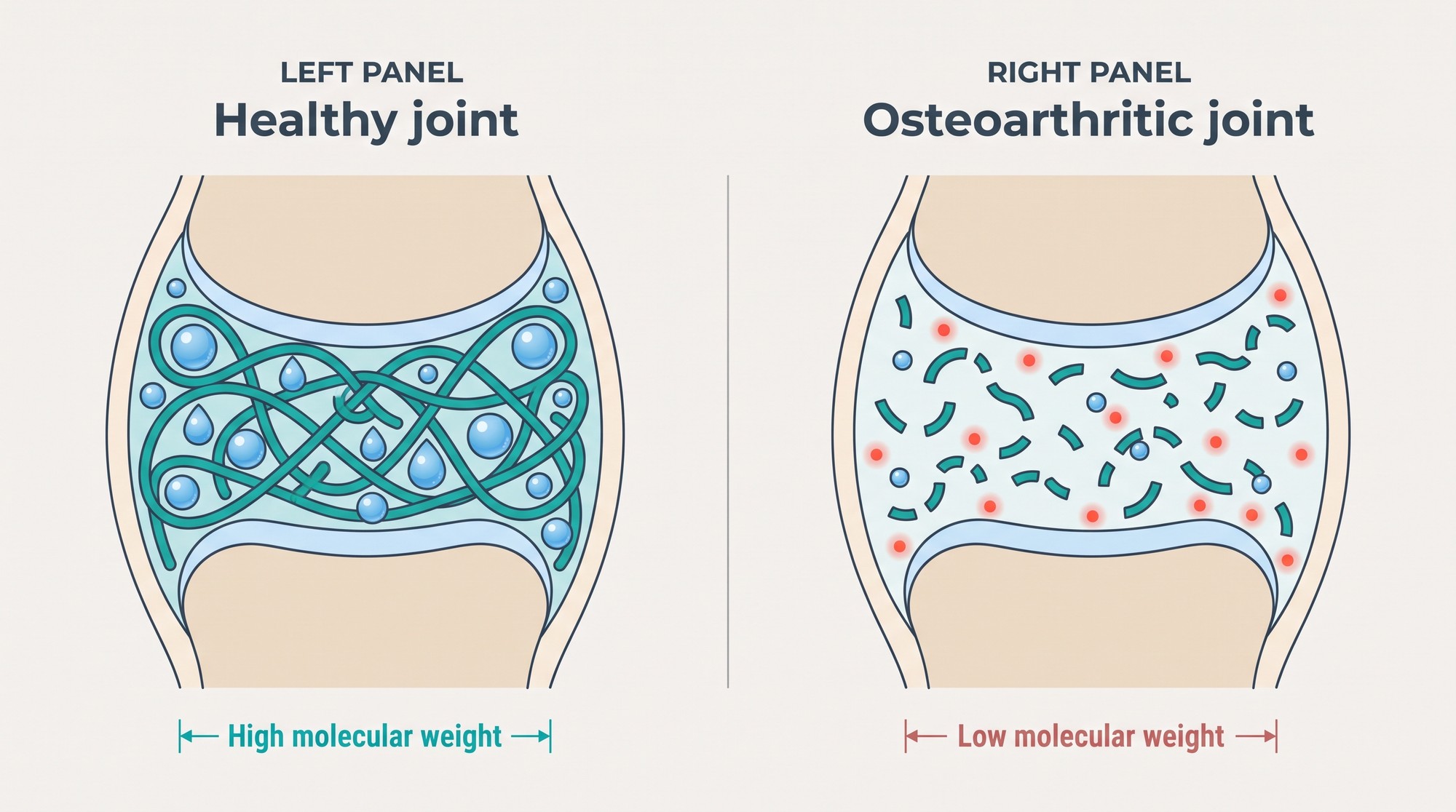
Knee osteoarthritis serves as the prime clinical example of this environment failing over time. The joint space is normally filled with a viscous, highly elastic liquid. This fluid acts as a dynamic shock absorber during high-impact activities like running and as a smooth lubricant during slow movements like walking. The primary molecule responsible for these properties is hyaluronan, a long-chain sugar polymer that traps water and helps create a thick, gel-like layer around the joint surfaces. The same compound appears across human biology, as a look at how hyaluronic acid supports skin, joints and eyes makes clear. As osteoarthritis progresses, the synovial fluid hyaluronan concentration and molecular weight fall, and the fluid becomes thinner and less able to disperse mechanical loads. It loses much of its capacity to cushion and protect the fragile cartilage surfaces, and mechanical wear gains the upper hand.
The Inflammatory Cascade
This physical damage triggers an aggressive biological response inside the joint space. Joint inflammation rises, along with a wave of cytokines, the signaling proteins that can instruct the body to break down local tissue. Interleukin-1 beta and tumor necrosis factor alpha increase in concentration, prompting local cells to release enzymes that degrade collagen fibers and undermine the structural integrity of the joint. The resulting environment is hostile to healthy cartilage. Breakdown products from the damaged cartilage float in the thinned fluid and irritate the joint lining, where they can provoke further inflammation.
Restoring the Mechanical Barrier
The medical response to this degraded environment often involves injecting a replacement fluid directly into the joint capsule, a procedure known as viscosupplementation. One established example is Hyalgan, a sterile, viscous solution of sodium hyaluronate derived from avian tissue. The preparation is intended to temporarily restore some of the physical properties of healthy joint fluid and provide direct mechanical support to the knee. Hyalgan is classified as a low-molecular-weight hyaluronic acid, a category covering preparations below roughly 1,500 kilodaltons; its stated range of 500 to 730 kilodaltons shapes how the injected material interacts with the cellular architecture inside the synovial space. It does not rebuild cartilage, and its role is generally described as symptom relief rather than structural repair. Clinics that buy Hyalgan for orthopedic clinics can consult the product listing for formulation and prescribing details.
Cellular Binding and Receptor Activation
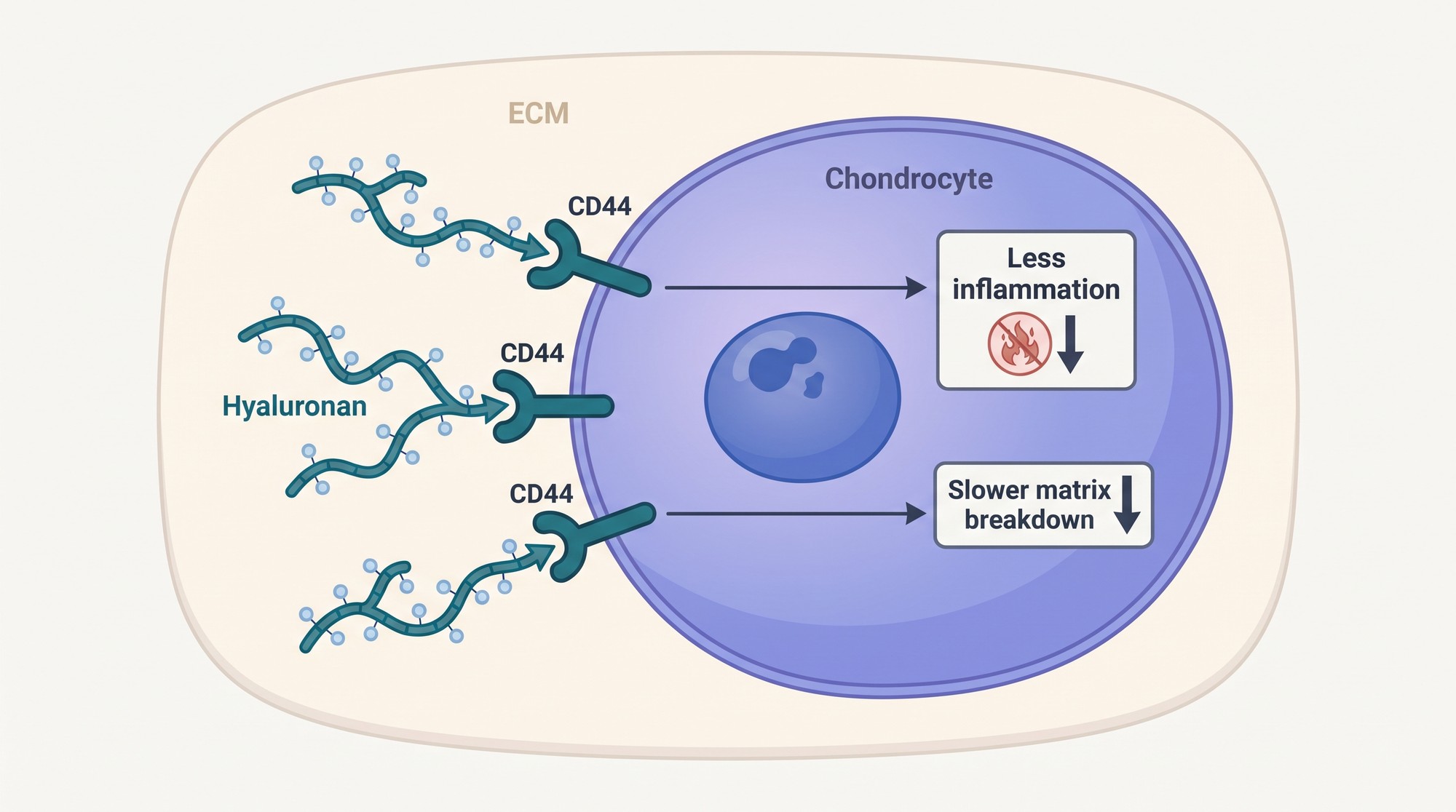
Intra-articular hyaluronic acid provides mechanical lubrication while also influencing the behavior of the cells that line and inhabit the joint. A 2015 systematic review of the mechanism of action found that binding to CD44 receptors on the surface of chondrocytes was the most frequently reported biological pathway in the basic science literature, with downstream effects that include reduced inflammatory signaling and less activity from the enzymes that break down cartilage matrix. Those receptors act as biological sensors that read the state of the joint fluid. Chondrocytes are the specialized cells responsible for building and maintaining the cartilage matrix. When they are stimulated by hyaluronan in this way, they tend to shift from a catabolic state toward one that favors matrix maintenance, a transition that is thought to slow cartilage degradation. The cells produce fewer of the metalloproteinases that degrade structural proteins and better stabilize their immediate environment.
Specific Biochemical Alterations
The biochemical shifts inside the joint capsule are generally described as following a common pattern. Evidence points to several alterations in the local environment after the introduction of the therapeutic fluid:
- A reduction in the concentration of pro-inflammatory mediators in the fluid space.
- An increase in the activity of natural enzyme inhibitors, which help protect the remaining collagen.
- Less mechanical load transferred to the subchondral bone as cushioning improves.
The local chemistry ultimately shapes the physical mechanics of the whole joint system.
Resetting the Synovial Membrane
The lining of the joint capsule also responds to the presence of the injected fluid. Synovial membrane function matters here because the membrane filters blood plasma and produces the fluid that lubricates the joint. In a diseased state, the inflamed membrane produces fewer and shorter hyaluronan molecules, which offer little mechanical protection. Injection of a concentrated formulation appears to interrupt that cycle, and the local cells respond by adjusting their own production.
The Production of Endogenous Hyaluronan
This response is often described as supporting endogenous hyaluronan synthesis, in which the joint begins producing higher-quality fluid again. The proposed explanation is that the injected material acts as a template for what the synovial tissue should be producing, which may help explain why clinical benefit is sometimes reported to outlast the injected gel itself. Exogenous hyaluronan is cleared from the joint relatively quickly, while symptom improvement can persist for months, although the exact relationship between the two is still debated. Over time the synovial tissue may move out of a state of chronic inflammation and back toward routine maintenance, with the joint space once again able to maintain boundary lubrication and defend itself against ordinary daily impact.
Clinical Execution and Timed Protocols
Treatment is delivered as a course rather than a single dose. Most orthopedic protocols rely on a timed series of sequential injections, and for low-molecular-weight preparations such as Hyalgan the labeled schedule is typically one injection per week for three to five consecutive weeks. Repeated exposure is intended to keep the injected material present in the joint long enough for the cellular response to develop while the joint adapts mechanically.
Tracking Functional Outcomes
Doctors monitor progress through standardized metrics to check whether the cellular changes translate into physical benefit. They track joint pain and function scores, which evaluate everything from resting pain levels to the ability to perform weight-bearing movements. The clinical goal is a measurable reduction in baseline pain and an improvement in daily mobility. Those scores offer an indirect reflection of what is happening inside the joint, and patients who respond to a full course often report an easier time with stairs or long walks and fewer inflammatory flare-ups.
Long-Term Joint Preservation
Cartilage cannot regrow once it is worn away down to the bone, so joint preservation depends on stopping the breakdown before bone is permanently exposed. Altering the composition of the synovial fluid is one of the more direct ways to influence the fate of the remaining viable tissue, because the chemical environment shapes the mechanics of the joint. Replacing degraded fluid with a viscosupplement gives the cells different mechanical signals to respond to and may encourage them to dial back catabolic activity.

