

While brain regions are known to activate during both sight and memory, a deeper mystery remained: do the exact same cells handle both tasks? New evidence suggests that seeing an object and picturing it in your mind often triggers the same individual neurons, revealing a shared neural code for perception and imagination. This breakthrough suggests that our internal imagination isn’t just a separate mental layer—it is a direct replay of the physical hardware we use to see the world.
Inside controlled clinical settings, researchers tracked single-neuron activity within brain areas dedicated to recognizing faces and objects. When participants closed their eyes to imagine what they had previously seen, a significant portion of those same neurons fired once more. This discovery provides rare, cell-level proof that the ‘mind’s eye’ isn’t just a metaphor; it’s a biological reality where your brain uses its own visual cortex code to simulate images without external light.
Think about the last time you mentally checked if you left the oven on or pictured a loved one’s smile. You weren’t just thinking in words—you were likely ‘seeing’ those images using the same neural architecture that processes your current surroundings. By listening to individual brain cells, this research helps us realize that our inner world and our outer reality are far more biologically connected than we ever realized.

Mapping the Shared Neural Code: Single-Neuron Evidence for The Mind’s Eye
Breakthrough in Brain Imaging: How Individual Neurons Handle Visualization
Neural systems assemble fleeting mental scenes from physical biological components, even if we experience these images as private internal broadcasts. This study is getting attention because it moves beyond brain scans and listens to individual cells.
This discovery is significant because the brain does not only “remember” with words. It frequently remembers with pictures. For example, your brain uses these codes when you replay a child’s face or picture exactly where you parked your car.
The research team analyzed over 700 individual neurons to map how our visual architecture handles imagination. By looking at these specific biological markers, they identified clear patterns in how the mind’s eye reactivates.
- Study published in Science on April 9, 2026.
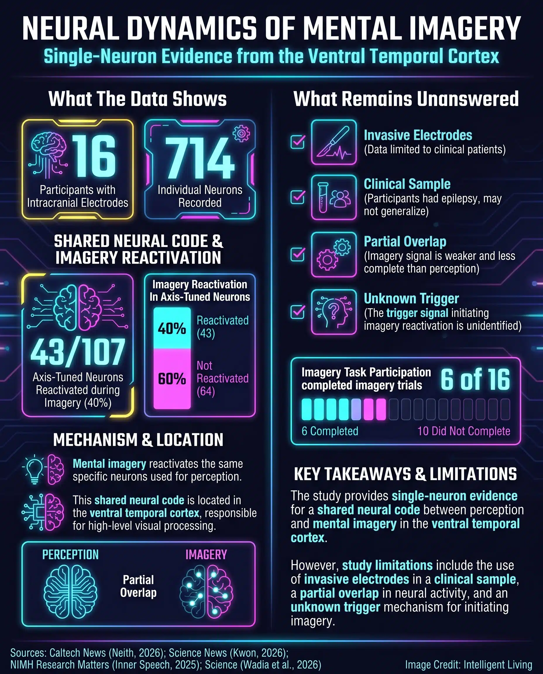
- Researchers recorded more than 700 individual neurons in the ventral temporal cortex, part of the ventral visual stream for object recognition that helps the brain tell faces and objects apart.
- Participants included 16 epilepsy patients undergoing clinical monitoring with implanted electrodes.
- Around 80 percent of visually responsive neurons followed a consistent “axis code” pattern for representing objects.
- Roughly 40 percent of relevant neurons reactivated during mental imagery tasks in a subset of participants.
These statistics suggest that imagination isn’t just a faint memory but a functional replay of our visual system. Such data explains why our mental pictures often feel remarkably grounded in reality.
The headline numbers are simple, but the story underneath is more subtle. This is not a claim that imagination is identical to vision. It is evidence that imagination can reuse some of vision’s working parts.

Scientific Results: How Individual Cells Reactivate During Imagination
Neural Mapping: Identifying the Visual Architecture of The Brain
Anatomical hubs like the ventral temporal cortex, located on the brain’s underside, drive our ability to recognize faces, places, and everyday objects. This area includes specialized parts of the fusiform gyrus linked to object selectivity that help the brain tell similar-looking things apart.
Replay Mechanisms: The Same Neural Circuitry Reappears During Imagination
Later, when those same individuals were asked to imagine some of those images, many of the same neurons fired again in similar configurations. Researchers found that imagining an object reactivates the very same neurons used to see it, bridging the gap between perception and the mind’s eye.
Picture a crowded airport: you recognize a friend from a split-second mental snapshot moments before they physically appear. This finding suggests the brain’s internal snapshot is a biological fact. It involves a partial replay of the same neural circuitry that processed the face initially.
Why Cell-Level Evidence Changes the Conversation
Cell-level overlap proves that the mind’s eye borrows tangible visual hardware, shifting the scientific consensus away from the idea of isolated imagination. This breakthrough gives researchers a sharper handle on what to measure next. Researchers are now able to track which parts of the visual code return during imagination and which specific circuits stay silent.
Experimental Design: Tracking Perception and Imagery in Real Time
Why Hospital Epilepsy Monitoring Makes this Possible
The study relied on a rare opportunity. Sixteen adults with epilepsy were already undergoing clinical monitoring in hospital settings. As part of their treatment, thin electrodes had been implanted to track seizure activity.
With consent, researchers used those electrodes to record signals from single neurons in the ventral temporal cortex. This type of single-neuron recording during epilepsy monitoring is one of the few ways to watch individual human brain cells in action while patients are already being observed for clinical reasons.
The Two-Phase Task: Seeing First, then Imagining
Initial phases involved participants viewing various objects while researchers measured the strength of their neuronal responses. During the following stage, these same individuals closed their eyes to imagine the images, allowing for a direct comparison of brain recording data gathered during the investigation:
- Scientists recorded over 700 neurons in the ventral temporal cortex.
- Roughly 450 of these neurons showed selectivity for certain visual categories.
- Approximately 40 percent of relevant neurons reactivated when participants closed their eyes to imagine specific items.
What “Decoding” Means in this Study
Computational models allowed researchers to decode these specific neural patterns with high accuracy. In controlled tasks, they were able to reconstruct which object a participant had seen or imagined at rates above chance. That does not mean thoughts were freely readable. It means that within a limited experimental setup, patterns in neural firing carried recognizable information.
Brainwave decoding into rough image sketches previously relied on scalp sensors, but implanted recordings now capture much more precise signals. The practical boundary remains: decoding depends on tight conditions, training data, and known targets.

Explaining Shared Neural Codes: A Simple Map of Your Brain’s Memory
A Pattern the Brain Reuses
Shared neural codes might sound complex, yet the underlying biological concept remains remarkably intuitive. When the brain sees an object, groups of neurons respond in a coordinated pattern. Each object produces a slightly different pattern, like a fingerprint made of electrical activity.
The Axis Code, Explained like a Map
Distributed ‘axis codes’ spread object details across vast cellular networks rather than isolating them in a single neuron. Researchers found that a distributed axis code maps object features across the visual system, with around 80 percent of visually responsive neurons fitting this pattern. Findings confirm that 80 percent of responsive neurons follow this specific mapping, while 40 percent reactivate during active imagination.
Axis-style mapping results echo earlier primate work where cells in the inferotemporal cortex functioned like sliders along core visual dimensions. The new study suggests a related kind of structure can also help explain what happens when the brain imagines.
What Reactivation Looks like in Plain Terms
When participants imagined an object, a portion of the original neural fingerprint reappeared. Although it was not a perfect replay and some cells remained silent, the overlap was unmistakable. The evidence shows that imagination taps directly into the visual system’s existing architecture.
Before revisiting a childhood home after years away, many people can mentally walk the hallway, “see” the kitchen light, and predict where the stairs turn. The evidence here suggests that this kind of mental walk-through is built from the same basic neural building blocks that originally processed walls, windows, and furniture.

Future Applications: Why these Neural Breakthroughs Matter for You
Basic science findings like these lead directly to questions we often ask ourselves, such as why some memories feel so vivid or how an imagined scene helps us plan and practice for the future.
The early answers are about circuits and codes, not quick fixes. Still, the overlap between vision and imagination provides a clearer target for researchers who study mental health, memory, and brain-computer interfaces.
Impact on Mental Health: Decoding Intrusive Images and Flashbacks
Unwanted scenes can feel intensely visual because imagination partially reuses our perception circuits. Such flashes often occur even when our eyes are closed or nothing is in front of us. Although this connection is not a treatment, it provides a clearer map for studying conditions where imagery and reality blur.
Stress systems significantly alter how the brain processes memory, particularly within circuits tied to visual imagery. Separate clinical discussions mention a neuroplasticity window in trauma recovery when the brain is more adaptable. This new finding allows scientists to test which parts of the visual code return during intrusive imagery.
Improving Brain-Computer Interface Research
Neural decoding breakthroughs prove that brain signals can be translated into text or imagery, creating new possibilities for human communication. Many brain-to-text systems still hit a decoder training bottleneck in brain-to-text systems that forces person-by-person calibration, which slows real clinical rollout.
Some implanted systems have already enabled BCI communication for locked-in syndrome by translating neural activity into letters, showing what is possible when hardware and training align. A better grasp of shared neural codes could refine those efforts, though real-world applications remain limited and highly technical.

Deepening Models of Memory and Learning
Biological stability in memory relies on mental imagery to ensure new learning doesn’t disrupt existing knowledge. The idea of a silent synapse reserve system in adult learning helps explain how new capacity can switch on without bulldozing older circuits.
Some teams also explore electrical neurostimulation to boost learning and memory to see whether timing and targeting can nudge recall in the right direction. As researchers get better at describing the “fingerprint” of an image in the brain, they can test whether stimulation strengthens useful recall or simply adds noise.
Clarifying Why Some People Struggle to Visualize
Aphantasia, defined as a difficulty forming mental pictures, highlights the variability in how neural codes reactivate. Some people report a blank screen when asked to picture a loved one’s face, while others describe a crisp, colorful image.
This study does not directly test aphantasia, but it helps frame what future experiments could measure. Instead of asking only how vivid a person’s imagery feels, researchers can ask whether fewer neurons reactivate, whether the pattern is weaker, or whether the brain uses a different route to build imagination.
Everyday Impact: Why Mental Rehearsal Can Work
Think about the last time you prepared for a big presentation by picturing the room and your audience. If the brain is reusing parts of the same visual code it uses for perception, that rehearsal effectively primes your neural pathways. This mental walk-through uses a shared neural code to prepare the system for real-world execution, making your actual performance feel more familiar and controlled.
What this Means and What it Still Can’t Do
What it Does Not Prove Yet
Why the Study Group Matters
Despite the excitement, several limits are clear. This unique research opportunity allows scientists to see how neurons follow the same electrical path during both real sight and mental imagery in a hospital setting.
The participants were epilepsy patients undergoing clinical monitoring, not a broad cross section of the population. That does not erase the value of the finding, but it does mean the results should be tested across more people and more brain locations when future methods allow.
The Missing Trigger Signal
Imagery strength varies widely from person to person. The study also leaves open a crucial question: what signal in the brain triggers the shift from passive memory to active mental imagery?
The researchers still need to understand how the brain distinguishes between something truly seen and something only imagined. The overlap is meaningful, but it is not total, and the brain usually keeps imagined scenes from being mistaken for real perception.
The Mind’s Eye and the Future of Brain Research
A Shared Code, Not A Shortcut
The discovery that the same neurons can help power both sight and imagination reframes a basic human experience. Seeing and picturing are not separate mental worlds. They are intertwined processes built from shared neural code.
For anyone who has replayed a memory before sleep or visualized a route before driving, the implication is striking. The brain is not inventing images from scratch. It is reusing its own visual hardware in ways that are measurable at the level of single cells.
Basic neural mechanisms often take years to translate into real-world medical interventions, meaning this cellular overlap is not yet a diagnostic tool.
What Researchers are Likely to Test Next
Upcoming research is expected to focus on the signals that trigger neural replay and how the brain distinguishes between imagination and reality. Those answers could improve scientific models of memory, sharpen how brain-computer interfaces are trained, and clarify why the mind’s eye is vivid for some people and muted for others.

Why Does the Brain Reuse Neurons for Sight and Imagination?
This discovery that individual neurons power both sight and imagination fundamentally changes how we view human memory. We now know that seeing and picturing are not separate mental activities, but rather intertwined processes built from a shared neural code. For anyone who has ever replayed a memory before sleep or visualized a route before driving, the implication is powerful: your brain isn’t inventing these images from scratch but rather reusing its own visual hardware in measurable, cell-level patterns.
Looking ahead, researchers want to uncover the specific signal that triggers this neural replay and how the brain distinguishes between an imagined scene and reality. Basic neural mechanisms often take years to translate into real-world medical interventions, meaning this cellular overlap is not yet a diagnostic tool.
These answers could sharpen how we train brain-computer interfaces and explain why the mind’s eye feels vivid for some while remaining muted for others. While this research doesn’t unlock instant therapies, it provides a clearer map for understanding how our internal snapshots help us learn, plan, and remember the world around us.
Common Questions About How Neurons Handle Sight and Imagination
Do the same brain cells fire when you see and imagine?
Yes, research shows that a portion of the same neurons active during visual perception reactivate when you imagine those same objects later.
Which part of the brain controls the mind’s eye?
The ventral temporal cortex is a key region, as it contains specialized neurons that help the brain recognize and mentally replay faces, places, and items.
Can scientists read my thoughts using this neural research?
No. Current decoding relies on specialized intracranial monitoring used for epilepsy care, meaning results are restricted to specific laboratory tasks.
Why do some people have a more vivid imagination than others?
Vividness may depend on the strength of neural reactivation, though researchers are still studying how individual brain chemistry affects the ‘mind’s eye.’
Does this study explain why I can’t see mental pictures?
Alternative approaches such as non-hallucinogenic neuroplasticity treatments explore different methods for improving brain flexibility.


