
Macular damage frequently distorts central vision in age-related macular degeneration. This damage initiates a difficult transition toward total central blindness, forcing patients to reconstruct how they perceive their environment. Science Corporation recently secured significant funding to accelerate access to the PRIMA retinal implant, a breakthrough BCI eye implant designed to restore functional sight. Integrating camera glasses with a miniaturized subretinal chip allows the PRIMA system to rebuild essential neural pathways for central sight. Current clinical testing aims to prove that these interfaces can offer a practical solution for late-stage dry AMD treatment where biological interventions have historically failed.
Recent clinical data now establishes a precise trajectory for families navigating this condition. Rebuilding a visual signal requires a precise blend of neurotechnology and surgical expertise. The following evaluation examines how PRIMA functions, explores the peer-reviewed evidence, and highlights the critical milestones patients should monitor over the coming months.

PRIMA Retinal Implant Overview: $230M Funding, How it Works, and Real-World Independence
Core Facts on PRIMA Technology and Geographic Atrophy Vision Restoration
- PRIMA is designed for central vision loss in geographic atrophy due to age-related macular degeneration.
- The system combines camera glasses with a wireless subretinal implant that converts light into retinal stimulation.
- PubMed clinical summary for the geographic atrophy retinal implant study provides 12-month clinical data for subretinal implant performance.
- Safety objectives and evaluation protocols remain publicly available through the official registry for the PRIMAvera trial.
- Science Corporation recently closed a Series C round to fund PRIMA commercialization and scale manufacturing, positioning the device for wider deployment.
Restoring Vision and Independence through BCI Eye Implant Technology
How PRIMA Rebuilds a Central Vision Signal
Categorizing PRIMA requires nuance: it functions as a brain-computer interface without ever contacting the brain directly. Retinal neural tissue already communicates using the brain’s visual language. PRIMA’s goal is to rebuild a usable signal at the point where the signal normally begins.
Digitizing visual input to replace degenerated photoreceptor roles involves a coordinated sequence:
- A high-resolution camera captures the surrounding environment.
- A processor translates visual data into a simplified, high-contrast pattern.
- Near-infrared light projects this pattern onto a tiny photovoltaic subretinal chip.
- The implant converts light into electrical stimulation to activate remaining retinal circuitry.
Generated pulses transmit visual information through the optic nerve, allowing the brain to process a restored sensory signal.
Why Independence Outcomes Matter
A low-vision occupational therapist described a moment that often gets overlooked in device headlines: a patient sitting at a kitchen table, slowly relearning the outline of large letters and then reading a short label without asking for help. Functional utility takes precedence over cinematic appeal. Integrating wireless subretinal chips with camera glasses allows some participants to regain enough functional vision for reading, which resonates beyond charts.
Patients increasingly adopt assistive technologies, specifically smart glasses designed for low-vision navigation that mitigate environmental obstacles in public spaces.
Hardware developments emphasize the role of AI-powered augmented reality eyewear in optimizing battery life for all-day vision assistance. The PRIMA system differentiates itself by restoring a visual signal for tasks where audio cues alone fail to provide sufficient detail.

PRIMA in the Global BCI Landscape: Clinical Evidence and Comparative Analysis
“Brain Implant” Headlines Miss the Point: Eye Implants and Neural Tissue
Why Retina-Based Interfaces Feel More Concrete
Describing PRIMA as a brain implant misrepresents its clinical application. It is a subretinal eye implant positioned directly beneath the tissue where photoreceptor degeneration has occurred, ensuring localized stimulation. The hardware utilizes a miniaturized 378-pixel photovoltaic array to translate infrared patterns into localized neural stimulation.
System reliance on 880 nm near-infrared projections wirelessly powers the subretinal sensors to map visual data to the macula. Data suggests that subretinal photovoltaic interfaces restore functional central vision without compromising existing peripheral acuity, which matters for geographic atrophy because many patients depend on eccentric fixation to use remaining vision.
Retina-First Versus Cortex-First Tradeoffs
Retina-first neurotechnology may reach patients sooner than various cortex-first strategies. Vitreoretinal surgeons already operate in the eye, and clinical pathways for retinal surgery, follow-up imaging, and rehabilitation are established in many health systems.
Establishing clinical transparency ensures patients understand the sensory nature of the interface. Families often express concern regarding the phrase “brain implant” but feel differently when they learn the device is placed in the eye to restore a sensory signal.
Various neural interfaces designed for speech decoding operate non-invasively, bypassing the need for direct cortical contact. While powerful, direct visual cortex stimulation remains a specialized alternative to retinal-based strategies due to scaling difficulties.
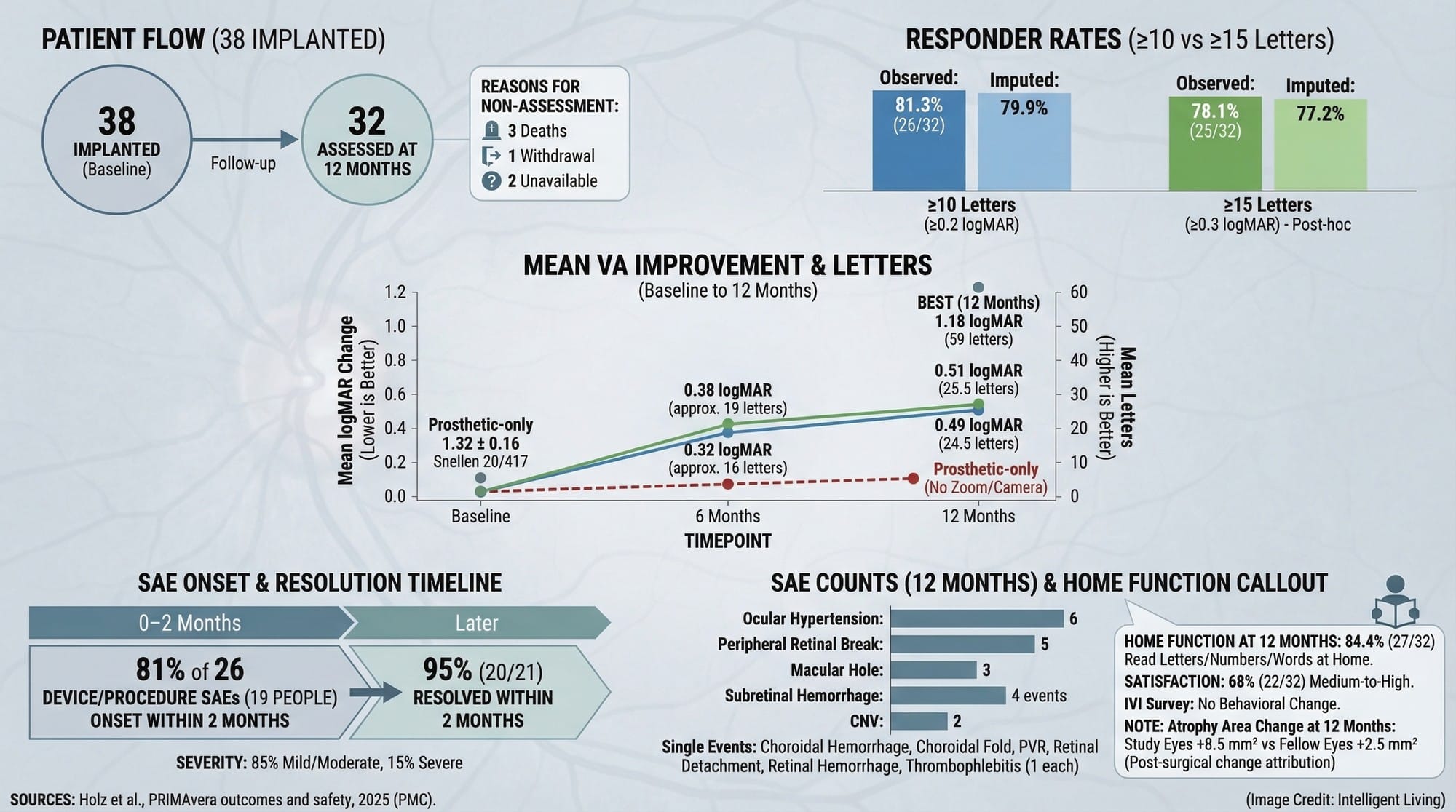
Analysis of NEJM Clinical Study Outcomes for Retinal Implants
Outcomes in Plain Numbers
Evidence for the device’s efficacy stems from a twelve-month clinical trial for vision restoration targeting advanced dry AMD. During that study, 38 participants received the implant, and 32 participants were assessed at 12 months. Integrating photovoltaic implants with specialized eyewear establishes a meaningful benchmark for improving acuity in degenerative conditions, which is a significant bar in a condition known for irreversible decline.
Impact assessment focuses on translating clinical tests into functional utility for daily living. Success is measured by whether patients can:
- Reliably identify letters and high-contrast patterns.
- Detect shapes in the center of their visual field.
- Perform real-world tasks such as reading large print without assistance.
Clinical staff members described a common scene during rehabilitation sessions: a person who has not read a printed label in years pointing to the correct line of text, then pausing, then doing it again with growing confidence. Autonomous function defines the true value of these outcomes, measured against years of progressive loss.
Safety Signals and Surgical Reality
Trial data indicates serious adverse events clustered immediately following surgery, though most resolved quickly. Significant risks exist. Every retinal implant requires both surgical precision and a long-term rehabilitation commitment, factors often complicated by the pre-existing health profiles of older AMD patients.
Study Design Limits and Confidence
While the open-label, single-group trial structure remains clinically valuable, the study design necessitates caution when generalizing outcomes across broader populations. Continuous evaluation increases the importance of longer follow-up, broader real-world datasets, and careful patient selection to clarify who benefits most.
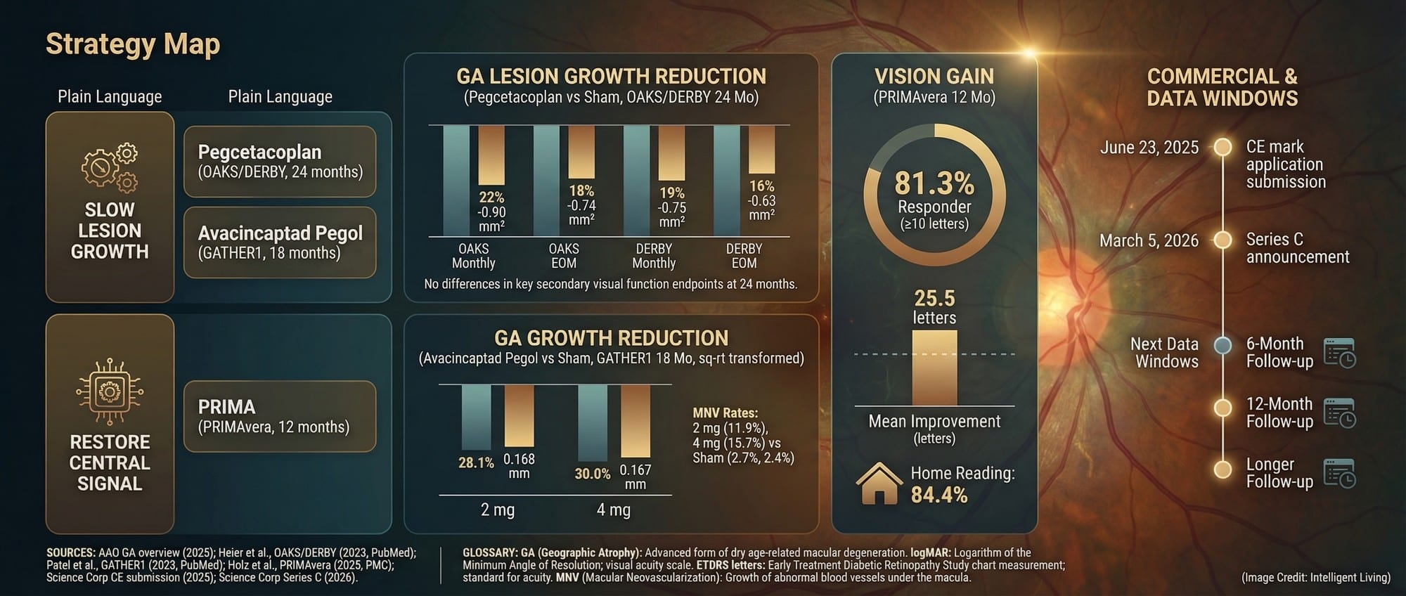
Geographic Atrophy Treatment Strategies and PRIMA Commercialization Milestones
Geographic Atrophy Treatment Dichotomy: Lesion Management Versus Functional Restoration
Slowing Lesion Growth is Not the Same as Restoring Vision
Ongoing destruction of the macula, as described in the American Academy of Ophthalmology geographic atrophy guide, creates a dense central blind spot. Macular tissue is responsible for detailed central vision, the part used for reading, faces, and fine tasks.
Recent medications focus on slowing progression, not restoring lost vision. Inhibiting damage spread preserves future function but fails to restore existing visual deficits.
Regulatory approvals for medications like pegcetacoplan focus on slowing the rate of GA growth rather than improving visual acuity or preventing visual acuity loss rather than reclaiming lost visual function, which is a different goal than signal restoration.
Where Neuroprosthetics Fit Alongside Regenerative Research
The PRIMA system is restorative in intent. The device does not reverse underlying degenerative biology. Stimulating remaining retinal circuitry rebuilds a usable signal. Individuals may one day face a choice between approaches, or a sequence of approaches, depending on disease stage.
Biological science offers several parallel paths for vision preservation:
- Recent breakthroughs in regenerative stem cell therapy explore the potential for producing visual gains by replacing degenerated tissue.
- Current pipelines for gene therapy and neural prosthetics address distinct failure points within the human visual system.
- Emerging nanotechnology for retinal protection focuses on delaying the timeline of vision loss at a cellular level.
Nanotechnology targets a specific point on the timeline of vision loss. The PRIMA system operates in the neuroprosthetic lane of signal restoration, while biological breakthroughs may eventually offer a tiered treatment roadmap for geographic atrophy.
Why $230M Now: Manufacturing, Training, and Access
Verified clinical data represents only the first milestone in the commercialization of medical devices. Commercial success depends on manufacturing precision, specialized surgeon training, and complex regulatory navigation. Analysts value the technology at approximately $1.5 billion following the recent Series C round, signaling high confidence in its path to commercialization.
What Scaling a Retinal Implant Actually Requires
Commercializing medical devices necessitates overcoming significant logistical hurdles beyond clinical trials. Scaling the PRIMA system involves:
- Maintaining extreme manufacturing precision for subretinal components.
- Implementing standardized surgeon training across diverse global health systems.
- Validating market confidence through robust investor participation.
Scheduled milestones will determine how quickly the PRIMA retinal implant reaches the general population.

Patient Action Steps and the Next 24 Months of Retinal BCI Progress
Practical Guidance for Families Managing Geographic Atrophy
Questions to Bring to a Retina Appointment
Current unavailability notwithstanding, families can adopt proactive measures to improve decision-making during the disease’s progression.
- Requesting a specialist explanation of disease stage and likely trajectory, including foveal involvement status.
- Seeking clear descriptions of treatment goals: slowing progression, restoring function, or supporting independence.
- Evaluating candidacy for future implant trials based on remaining retinal structure and overall eye health.
- Developing a low-vision plan now, including mobility, lighting, contrast, and daily task adaptations.
Support Tools that Preserve Independence Now
Proactive questioning transforms clinic visits into actionable decision points for families managing progressive vision loss. Proactive strategies include:
- Maintaining a list of questions regarding treatments that slow geographic atrophy.
- Identifying technologies capable of restoring central vision.
- Assessing how camera glasses support low-vision navigation.
Vascular BCI paths include the Stentrode, which reaches neural tissue through blood vessels rather than open surgery. EEG headphones that map attention and cognitive workload demonstrate how brain-signal mapping functions without surgical intervention.
Optional Future Horizon: Biohybrid Interfaces and the “Living Neuron” Bet
Biohybrid Interfaces: A Longer-Horizon Bet
Current PRIMA development focuses on providing a practical retinal neuroprosthetic for immediate clinical application. neural interface that integrate living neurons with electronics aim to integrate living neurons with circuitry for more natural signal transfer. Preclinical research explores optogenetic stimulation in biohybrid implants to determine if living tissue can improve information flow.
Neural Data, Neurorights, and Mental Privacy
Scaling interfaces increases the importance of governance. International ethics debates increasingly prioritize neurorights and cognitive privacy frameworks as essential safeguards for autonomous neural technology.
Critical Milestones for Retinal BCI Availability and Real-World Access
Several milestones will clarify whether PRIMA becomes a broadly available option or remains limited to specialized programs.
- Regulatory momentum is building following the submission of a European CE mark application for the PRIMA system.
- Projections indicate a rapid transition from testing to market availability for the commercial rollout of the retinal implant.
- Upgrading subretinal photovoltaic implants can improve prosthetic visual acuity significantly and enhance the resolution of prosthetic visual acuity.
- Retina specialists prioritize emerging therapies for geographic atrophy as surgical approaches move toward clinical consensus.
- Comparing decoder training highlights the significant learning curve required for sensory interfaces. Much like how decoder training bottlenecks speech BCI performance, vision implants rely on the patient’s neurological ability to interpret a restored signal.

Scaling Vision Restoration: The Future of PRIMA and Geographic Atrophy
The system shifts BCI development from experimental theory into measurable clinical application. Rebuilding a visual signal for a specific disability using established surgical pathways defines the current trajectory. Success relies on the synergy of manufacturing precision, durable patient outcomes, and robust rehabilitation support.
Independence gains for those with advanced macular degeneration are increasingly viable. Once technological development and operational infrastructure synchronize, the human impact becomes immediate and profound. Restoring visual signals allows a grandparent to recognize a grandchild’s smile or a person to read a simple label without assistance.
Common Questions About Vision Restoration and the PRIMA Implant
1. What is geographic atrophy in simple terms?
Progressive macular tissue death, as detailed in the BrightFocus geographic atrophy fact sheet, creates permanent blind spots in the center of vision.
2. How does the PRIMA subretinal chip restore sight?
The PRIMA subretinal chip converts near-infrared light from camera glasses into electrical pulses. These pulses stimulate remaining retinal circuitry, sending a visual signal to the brain that replaces lost photoreceptor function.
3. Is PRIMA considered a brain-computer interface?
Yes, PRIMA is a BCI eye implant. It interfaces directly with neural tissue in the retina—which is an extension of the brain—to restore sensory data without requiring surgery on the visual cortex.
4. Who can benefit from Science Corporation’s technology?
The technology currently targets patients with geographic atrophy due to AMD. It is designed for those who have lost central vision but still possess functional retinal layers capable of receiving electrical stimulation.
5. How does PRIMA differ from standard GA injections?
Standard injections focus on slowing the expansion of atrophy lesions to preserve remaining sight. PRIMA is a restorative approach that aims to bring back functional vision that has already been lost to the disease.


